Mazio Federica, Aloj Giuseppina, Pastorino Grazia Maria Giovanna, Perillo Teresa, Russo Carmela, Riccio Maria Pia, Covelli Eugenio Maria, Parasole Rosanna, Tedeschi Enrico, Ugga Lorenzo, D'Amico Alessandra, Quarantelli Mario
Pediatric Neuroradiology, Department of Neuroscience, Santobono-Pausilipon Children's Hospital, 80129 Naples, Italy.
Department of Pediatric Hemato-Oncology, A.O.R.N. Santobono-Pausilipon, 80123 Naples, Italy.
Biology (Basel). 2022 Mar 24;11(4):499. doi: 10.3390/biology11040499.
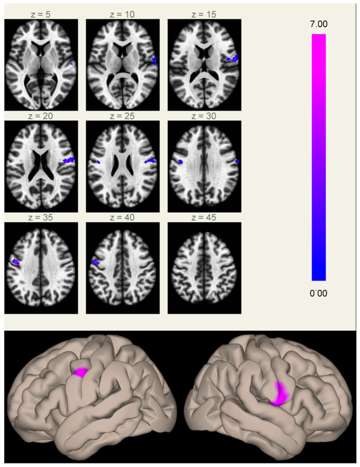
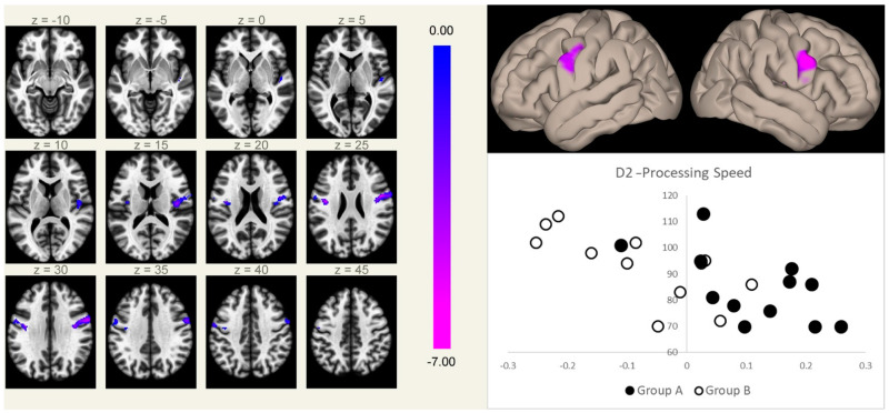
Whether chemotherapy (ChT) and radiotherapy (RT) determine neurocognitive impairment in acute lymphoblastic leukemia long-term survivors (ALL LTSs) through similar mechanisms affecting the same brain regions is still unknown. We compared neurocognitive alterations, regional brain tissue volumes (by voxel-based morphometry), and functional connectivity of the main default-mode network hubs (by seed-based analysis of resting state functional MRI data), in 13 ALL LTSs treated with RT and ChT (Group A) and 13 treated with ChT only (Group B). Group A performed significantly worse than Group B at the digit span and digit symbol tests ( = 0.023 and 0.013, respectively). Increased connectivity between the medial prefrontal cortex (the main anterior hub of the default-mode network) and the rolandic operculi was present in Group A compared to Group B, along with the absence of significant differences in regional brain tissue volumes. In these regions, the functional connectivity correlated inversely with the speed of processing scores, independent of treatment group. These results suggest that similar mechanisms may be involved in the neurocognitive deficits in ALL LTS patients, regardless of the treatment group. Further studies are needed to clarify whether these changes represent a direct expression of the mechanisms underlying the cognitive deficits or ineffective compensatory phenomena.
化疗(ChT)和放疗(RT)是否通过影响相同脑区的相似机制导致急性淋巴细胞白血病长期存活者(ALL LTSs)出现神经认知障碍,目前仍不清楚。我们比较了13例接受放疗和化疗的ALL LTSs(A组)和13例仅接受化疗的ALL LTSs(B组)的神经认知改变、区域脑组织体积(通过基于体素的形态学测量)以及主要默认模式网络枢纽的功能连接性(通过基于静息态功能磁共振成像数据的种子点分析)。在数字广度和数字符号测试中,A组的表现明显比B组差(分别为P = 0.023和0.013)。与B组相比,A组内侧前额叶皮质(默认模式网络的主要前枢纽)与中央 operculi之间的连接性增加,同时区域脑组织体积无显著差异。在这些区域,功能连接性与处理速度得分呈负相关,与治疗组无关。这些结果表明,无论治疗组如何,ALL LTS患者的神经认知缺陷可能涉及相似的机制。需要进一步研究来阐明这些变化是否代表认知缺陷潜在机制的直接表现或无效的代偿现象。