Hristov Bozhidar, Andonov Vladimir, Doykov Daniel, Tsvetkova Silvia, Doykova Katya, Doykov Mladen
Second Department of Internal Diseases, Section "Gastroenterology", Medical Faculty, Medical University of Plovdiv, 6000 Plovdiv, Bulgaria.
Gastroenterology Clinic, University Hospital "Kaspela", 4001 Plovdiv, Bulgaria.
Diagnostics (Basel). 2022 Mar 29;12(4):841. doi: 10.3390/diagnostics12040841.
A variety of imaging techniques exists for the diagnosis of pancreatic disorders. None of the broadly applied diagnostic methods utilizes elasticity as an indicator of tissue damage. A well-known fact is that both chronic pancreatitis (CP) and pancreatic ductal adenocarcinoma (PDA) are associated with the development of prominent fibrosis (increased tissue stiffness).
To prospectively assess the accuracy of point shear wave elastography (pSWE) in differentiating between benign and malignant pancreatic diseases, establish a cut-off value for the diagnosis of PDA, and evaluate the influence of certain variables on the obtained results.
The present study included 78 patients who were admitted at the Department of Gastroenterology at the university hospital "Kaspela" between December 2017 and August 2021 for diagnosis and/or treatment of pancreatic disorders. Based on the clinical criteria, diagnostic imaging, and histological findings, patients were divided into the CP and PDA group. The ultrasound based pSWE technique was applied and shear wave velocity (SWV) was measured. The depth of region of interest (ROI) and successful measurement rate were also recorded.
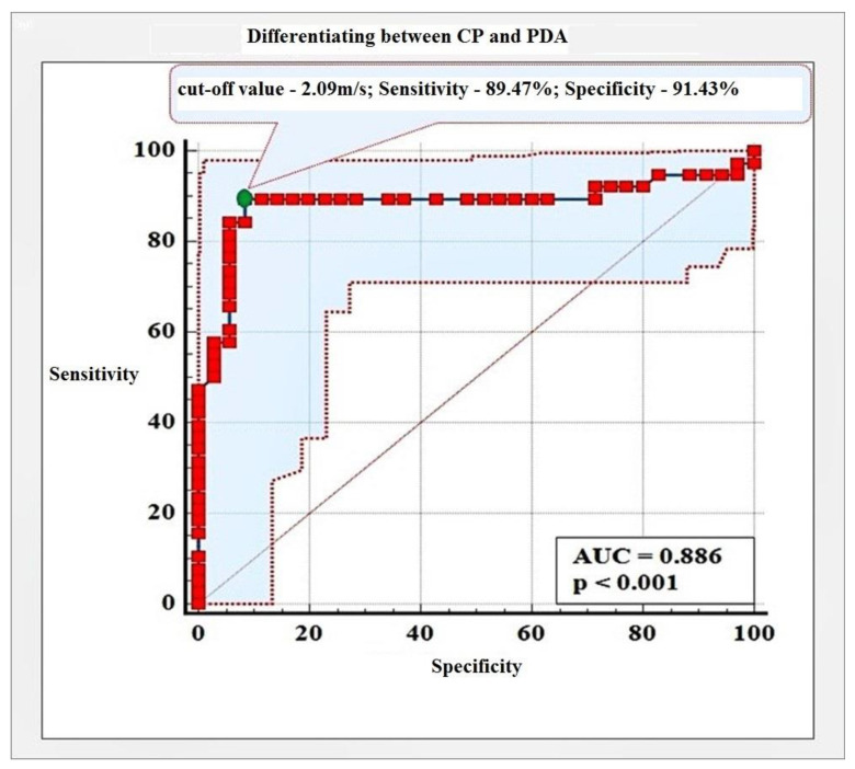
The mean ± SD SWV values established through pSWE were 1.75 ± 0.34 m/s and 2.93 ± 0.91 m/s for the CP and PDA, respectively. With a cut-off value of 2.09 m/s, we calculated the sensitivity (Se), specificity (Sp), and accuracy for differentiating between CP and PDA of 89.47%, 91.20%, and 88.60%, respectively. Of the examined variables, BMI and depth of ROI in the CP group and sex in the PDA group showed a statistically significant influence on the obtained results.
pSWE may be utilized as a differential diagnostic modality in patients with suspected CP or PDA.
存在多种用于诊断胰腺疾病的成像技术。广泛应用的诊断方法均未将弹性作为组织损伤的指标。一个众所周知的事实是,慢性胰腺炎(CP)和胰腺导管腺癌(PDA)均与显著纤维化(组织硬度增加)的发展相关。
前瞻性评估点剪切波弹性成像(pSWE)在鉴别良性和恶性胰腺疾病方面的准确性,确定PDA诊断的临界值,并评估某些变量对所得结果的影响。
本研究纳入了2017年12月至2021年8月期间在大学医院“卡斯佩拉”胃肠病科因胰腺疾病诊断和/或治疗而入院的78例患者。根据临床标准、诊断性成像和组织学检查结果,将患者分为CP组和PDA组。应用基于超声的pSWE技术并测量剪切波速度(SWV)。还记录了感兴趣区域(ROI)的深度和成功测量率。
通过pSWE确定的CP组和PDA组的平均±标准差SWV值分别为1.75±0.34米/秒和2.93±0.91米/秒。以2.09米/秒为临界值,我们计算出鉴别CP和PDA的敏感性(Se)、特异性(Sp)和准确性分别为89.47%、91.20%和88.60%。在所检查的变量中,CP组的BMI和ROI深度以及PDA组的性别对所得结果显示出统计学上的显著影响。
pSWE可作为疑似CP或PDA患者的鉴别诊断方法。