From Neurology (L.G., T.C., N.M.-C., M.M.-K.), Clinical Neurophysiology (S.B., E.W.), Cardiology (J.D.), and Anaesthesia and Intensive Care (H.F.), Department of Clinical Sciences Lund, Lund University, Skåne University Hospital, Malmö; Department of Psychiatry and Neurochemistry (K.B., H.Z.), Institute of Neuroscience and Physiology, the Sahlgrenska Academy, University of Gothenburg; Clinical Neurochemistry Laboratory (K.B., H.Z.), Sahlgrenska University Hospital, Mölndal, Sweden; Department of Cardiology (C.H.), Rigshospitalet and Department of Clinical Medicine, University of Copenhagen, Denmark; Departments of Intensive Care (J.H.) and Neurology/Clinical Neurophysiology (A.-F-V.R.), Amsterdam Neuroscience, Amsterdam UMC, Academic Medical Center, University of Amsterdam, the Netherlands; Departments of Clinical Neurophysiology (T.W.K.) and Cardiology (J.K.), Rigshospitalet University Hospital, Copenhagen, Denmark; Department of Intensive Care (M.K.), Medical Center Leeuwarden, the Netherlands; Clinical Memory Research Unit, Faculty of Medicine (N.M.-C.), and Wallenberg Centre for Molecular Medicine (N.M.-C.), Lund University; Anaesthesia and Intensive Care, Department of Clinical Sciences Lund (N.N.), Lund University, Helsingborg Hospital, Sweden; Department of Neurology (A.O.R.), CHUV and University of Lausanne, Switzerland; Department of Anesthesia and Intensive Care (P.S.), Centre Hospitalier de Luxembourg; Department of Life Sciences and Medicine (P.S.), Faculty of Science, Technology and Medicine, University of Luxembourg; Clinical Studies Sweden (S.U.), Skåne University Hospital, Lund; Department of Neurodegenerative Disease (H.Z.), UCL Institute of Neurology; UK Dementia Research Institute at UCL (H.Z.), London, UK; and Hong Kong Center for Neurodegenerative Diseases (H.Z.), China.
Neurology. 2022 Jun 14;98(24):e2487-e2498. doi: 10.1212/WNL.0000000000200335. Epub 2022 Apr 25.
EEG is widely used for prediction of neurologic outcome after cardiac arrest. To better understand the relationship between EEG and neuronal injury, we explored the association between EEG and neurofilament light (NfL) as a marker of neuroaxonal injury, evaluated whether highly malignant EEG patterns are reflected by high NfL levels, and explored the association of EEG backgrounds and EEG discharges with NfL.
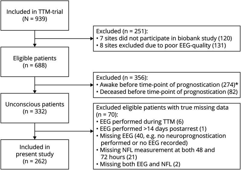
We performed a post hoc analysis of the Target Temperature Management After Out-of-Hospital Cardiac Arrest trial. Routine EEGs were prospectively performed after the temperature intervention ≥36 hours postarrest. Patients who awoke or died prior to 36 hours postarrest were excluded. EEG experts blinded to clinical information classified EEG background, amount of discharges, and highly malignant EEG patterns according to the standardized American Clinical Neurophysiology Society terminology. Prospectively collected serum samples were analyzed for NfL after trial completion. The highest available concentration at 48 or 72 hours postarrest was used.
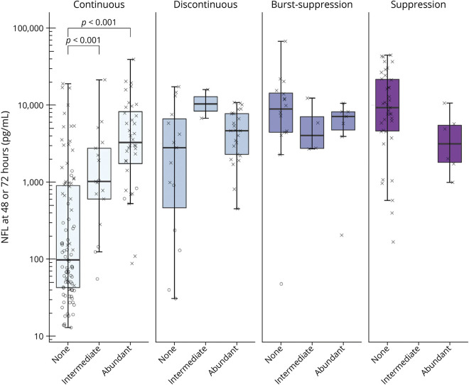
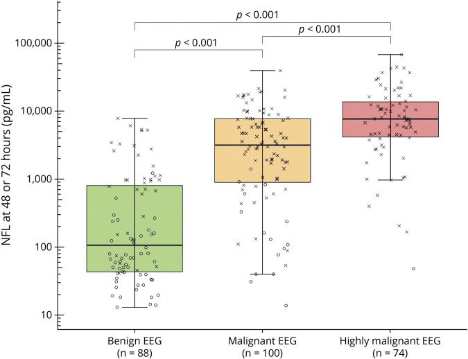
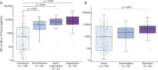
A total of 262/939 patients with EEG and NfL data were included. Patients with highly malignant EEG patterns had 2.9 times higher NfL levels than patients with malignant patterns and NfL levels were 13 times higher in patients with malignant patterns than those with benign patterns (95% CI 1.4-6.1 and 6.5-26.2, respectively; effect size 0.47; < 0.001). Both background and the amount of discharges were independently strongly associated with NfL levels ( < 0.001). The EEG background had a stronger association with NfL levels than EEG discharges (R = 0.30 and R = 0.10, respectively). NfL levels in patients with a continuous background were lower than for any other background (95% CI for discontinuous, burst-suppression, and suppression, respectively: 2.26-18.06, 3.91-41.71, and 5.74-41.74; effect size 0.30; < 0.001 for all). NfL levels did not differ between suppression and burst suppression. Superimposed discharges were only associated with higher NfL levels if the EEG background was continuous.
Benign, malignant, and highly malignant EEG patterns reflect the extent of brain injury as measured by NfL in serum. The extent of brain injury is more strongly related to the EEG background than superimposed discharges. Combining EEG and NfL may be useful to better identify patients misclassified by single methods.
ClinicalTrials.gov NCT01020916.
EEG 广泛用于预测心脏骤停后的神经功能预后。为了更好地理解 EEG 与神经元损伤之间的关系,我们探讨了 EEG 与神经丝轻链(NfL)之间的关系,NfL 是神经轴突损伤的标志物,评估高度恶性 EEG 模式是否反映在高 NfL 水平上,并探讨 EEG 背景和 EEG 放电与 NfL 的关系。
我们对心脏骤停后目标温度管理试验进行了事后分析。在体温干预后≥36 小时,常规进行 EEG。排除在 36 小时后苏醒或死亡的患者。EEG 专家根据标准化的美国临床神经生理学会术语,对 EEG 背景、放电量和高度恶性 EEG 模式进行盲法分类。在试验完成后,分析前瞻性采集的血清样本中的 NfL。使用 48 或 72 小时后最高的可用浓度。
共纳入 262/939 例有 EEG 和 NfL 数据的患者。高度恶性 EEG 模式患者的 NfL 水平比恶性模式患者高 2.9 倍,恶性模式患者的 NfL 水平比良性模式患者高 13 倍(95%CI 1.4-6.1 和 6.5-26.2,分别;效应大小 0.47;<0.001)。EEG 背景和放电量均与 NfL 水平独立高度相关(<0.001)。EEG 背景与 NfL 水平的相关性强于 EEG 放电(R=0.30 和 R=0.10)。连续 EEG 背景患者的 NfL 水平低于其他任何背景(不连续、爆发抑制和抑制的 95%CI 分别为:2.26-18.06、3.91-41.71 和 5.74-41.74;效应大小 0.30;均<0.001)。抑制与爆发抑制之间的 NfL 水平无差异。如果 EEG 背景连续,叠加放电仅与更高的 NfL 水平相关。
良性、恶性和高度恶性 EEG 模式反映了血清中 NfL 测量的脑损伤程度。脑损伤程度与 EEG 背景的关系比叠加放电更为密切。结合 EEG 和 NfL 可能有助于更好地识别单方法分类错误的患者。
ClinicalTrials.gov NCT01020916。