• 文献检索
  • 文档翻译
  • 深度研究
  • 学术资讯
  • Suppr Zotero 插件Zotero 插件
  • 邀请有礼
  • 套餐&价格
  • 历史记录
应用&插件
Suppr Zotero 插件Zotero 插件浏览器插件Mac 客户端Windows 客户端微信小程序
定价
高级版会员购买积分包购买API积分包
服务
文献检索文档翻译深度研究API 文档MCP 服务
关于我们
关于 Suppr公司介绍联系我们用户协议隐私条款
关注我们

Suppr 超能文献

核心技术专利:CN118964589B侵权必究
粤ICP备2023148730 号-1Suppr @ 2026

文献检索

告别复杂PubMed语法,用中文像聊天一样搜索,搜遍4000万医学文献。AI智能推荐,让科研检索更轻松。

立即免费搜索

文件翻译

保留排版,准确专业,支持PDF/Word/PPT等文件格式,支持 12+语言互译。

免费翻译文档

深度研究

AI帮你快速写综述,25分钟生成高质量综述,智能提取关键信息,辅助科研写作。

立即免费体验

磁共振成像中的新型材料:高介电常数陶瓷、超材料、超表面和人工电介质。

Novel materials in magnetic resonance imaging: high permittivity ceramics, metamaterials, metasurfaces and artificial dielectrics.

机构信息

Department of Radiology, C.J. Gorter MRI Centre, Leiden University Medical Center, Leiden, The Netherlands.

School of Physics and Engineering, ITMO University, Saint Petersburg, Russia.

出版信息

MAGMA. 2022 Dec;35(6):875-894. doi: 10.1007/s10334-022-01007-5. Epub 2022 Apr 26.

DOI:10.1007/s10334-022-01007-5
PMID:35471464
原文链接:https://pmc.ncbi.nlm.nih.gov/articles/PMC9596558/
Abstract

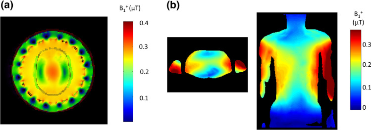
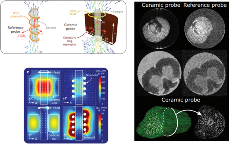
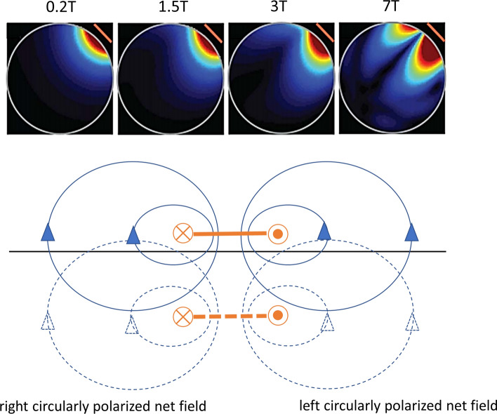
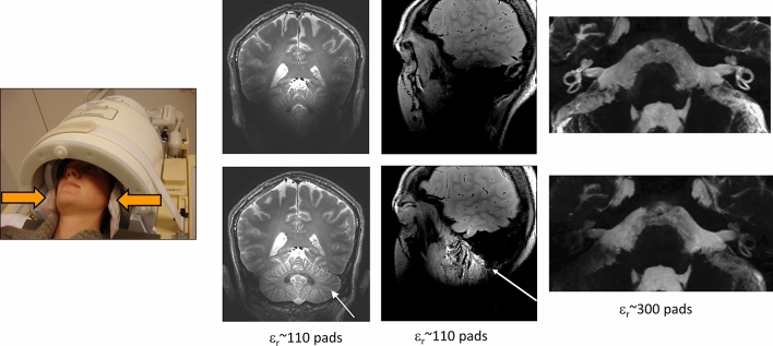
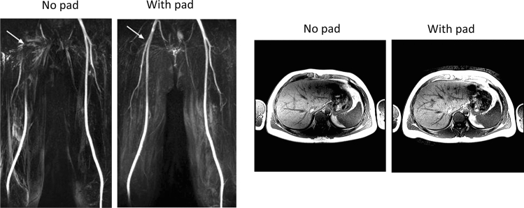
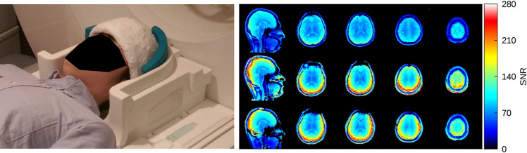
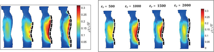
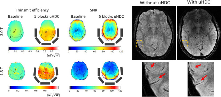
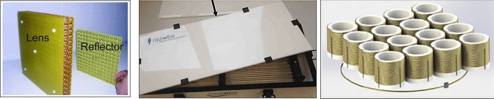
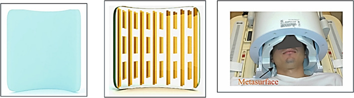
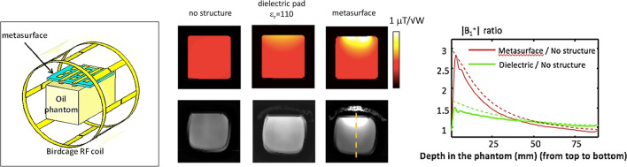
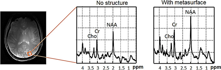
This article reviews recent developments in designing and testing new types of materials which can be: (i) placed around the body for in vivo imaging, (ii) be integrated into a conventional RF coil, or (iii) form the resonator itself. These materials can improve the quality of MRI scans for both in vivo and magnetic resonance microscopy applications. The methodological section covers the basic operation and design of two different types of materials, namely high permittivity materials constructed from ceramics and artificial dielectrics/metasurfaces formed by coupled conductive subunits, either in air or surrounded by dielectric material. Applications of high permittivity materials and metasurfaces placed next to the body to neuroimaging and extremity imaging at 7 T, body and neuroimaging at 3 T, and extremity imaging at 1.5 T are shown. Results using ceramic resonators for both high field in vivo imaging and magnetic resonance microscopy are also shown. The development of new materials to improve MR image quality remains an active area of research, but has not yet found significant use in clinical applications. This is mainly due to practical issues such as specific absorption rate modelling, accurate and reproducible placement, and acceptable size/weight of such materials. The most successful area has been simple "dielectric pads" for neuroimaging at 7 T which were initially developed somewhat as a stop-gap while parallel transmit technology was being developed, but have continued to be used at many sites. Some of these issues can potentially be overcome using much lighter metasurfaces and artificial dielectrics, which are just beginning to be assessed.

摘要

本文综述了设计和测试新型材料的最新进展,这些新型材料可以:(i)置于体内进行体内成像,(ii)集成到常规射频线圈中,或(iii)形成谐振器本身。这些材料可以提高体内和磁共振显微镜应用的 MRI 扫描质量。方法部分涵盖了两种不同类型材料的基本操作和设计,即由陶瓷制成的高介电常数材料和由耦合导电子单元形成的人工介电常数/超材料,无论是在空气中还是在介电材料周围。展示了高介电常数材料和超材料应用于神经成像和 7T 四肢成像、3T 身体和神经成像以及 1.5T 四肢成像的情况。还展示了陶瓷谐振器在高场体内成像和磁共振显微镜中的应用结果。改善磁共振图像质量的新材料的发展仍然是一个活跃的研究领域,但尚未在临床应用中得到广泛应用。这主要是由于实际问题,如比吸收率建模、准确和可重复的放置以及此类材料的可接受尺寸/重量。最成功的领域是 7T 神经成像的简单“介电垫”,最初是在并行传输技术开发的同时作为权宜之计开发的,但在许多站点仍在继续使用。使用更轻的超材料和人工介电常数可以潜在地克服其中一些问题,这些问题才刚刚开始评估。

https://cdn.ncbi.nlm.nih.gov/pmc/blobs/81fb/9596558/dcb2cc63aa18/10334_2022_1007_Fig24_HTML.jpg
https://cdn.ncbi.nlm.nih.gov/pmc/blobs/81fb/9596558/fa930b8ea387/10334_2022_1007_Fig1_HTML.jpg
https://cdn.ncbi.nlm.nih.gov/pmc/blobs/81fb/9596558/8169f4a9de54/10334_2022_1007_Fig2_HTML.jpg
https://cdn.ncbi.nlm.nih.gov/pmc/blobs/81fb/9596558/0543cde68828/10334_2022_1007_Fig3_HTML.jpg
https://cdn.ncbi.nlm.nih.gov/pmc/blobs/81fb/9596558/c5babaad5994/10334_2022_1007_Fig4_HTML.jpg
https://cdn.ncbi.nlm.nih.gov/pmc/blobs/81fb/9596558/953ddfdf1144/10334_2022_1007_Fig5_HTML.jpg
https://cdn.ncbi.nlm.nih.gov/pmc/blobs/81fb/9596558/4c5596c4f610/10334_2022_1007_Fig6_HTML.jpg
https://cdn.ncbi.nlm.nih.gov/pmc/blobs/81fb/9596558/106f2538db9e/10334_2022_1007_Fig7_HTML.jpg
https://cdn.ncbi.nlm.nih.gov/pmc/blobs/81fb/9596558/1a401c9a2e25/10334_2022_1007_Fig8_HTML.jpg
https://cdn.ncbi.nlm.nih.gov/pmc/blobs/81fb/9596558/f02e1d8a1e1d/10334_2022_1007_Fig9_HTML.jpg
https://cdn.ncbi.nlm.nih.gov/pmc/blobs/81fb/9596558/b2fea8e83ca1/10334_2022_1007_Fig10_HTML.jpg
https://cdn.ncbi.nlm.nih.gov/pmc/blobs/81fb/9596558/f9c46539b6dc/10334_2022_1007_Fig11_HTML.jpg
https://cdn.ncbi.nlm.nih.gov/pmc/blobs/81fb/9596558/ec03eaef532d/10334_2022_1007_Fig12_HTML.jpg
https://cdn.ncbi.nlm.nih.gov/pmc/blobs/81fb/9596558/fa5ce304c8be/10334_2022_1007_Fig13_HTML.jpg
https://cdn.ncbi.nlm.nih.gov/pmc/blobs/81fb/9596558/f63530305eeb/10334_2022_1007_Fig14_HTML.jpg
https://cdn.ncbi.nlm.nih.gov/pmc/blobs/81fb/9596558/dfabdef272c0/10334_2022_1007_Fig15_HTML.jpg
https://cdn.ncbi.nlm.nih.gov/pmc/blobs/81fb/9596558/22d3af2862df/10334_2022_1007_Fig16_HTML.jpg
https://cdn.ncbi.nlm.nih.gov/pmc/blobs/81fb/9596558/cb3d9a7316d4/10334_2022_1007_Fig17_HTML.jpg
https://cdn.ncbi.nlm.nih.gov/pmc/blobs/81fb/9596558/1b928440a526/10334_2022_1007_Fig18_HTML.jpg
https://cdn.ncbi.nlm.nih.gov/pmc/blobs/81fb/9596558/ffb3ed26e713/10334_2022_1007_Fig19_HTML.jpg
https://cdn.ncbi.nlm.nih.gov/pmc/blobs/81fb/9596558/444cb029ebd1/10334_2022_1007_Fig20_HTML.jpg
https://cdn.ncbi.nlm.nih.gov/pmc/blobs/81fb/9596558/736c329d97c4/10334_2022_1007_Fig21_HTML.jpg
https://cdn.ncbi.nlm.nih.gov/pmc/blobs/81fb/9596558/023ae1bf0e94/10334_2022_1007_Fig22_HTML.jpg
https://cdn.ncbi.nlm.nih.gov/pmc/blobs/81fb/9596558/e7415d3e2e6d/10334_2022_1007_Fig23_HTML.jpg
https://cdn.ncbi.nlm.nih.gov/pmc/blobs/81fb/9596558/dcb2cc63aa18/10334_2022_1007_Fig24_HTML.jpg

相似文献

1
Novel materials in magnetic resonance imaging: high permittivity ceramics, metamaterials, metasurfaces and artificial dielectrics.磁共振成像中的新型材料:高介电常数陶瓷、超材料、超表面和人工电介质。
MAGMA. 2022 Dec;35(6):875-894. doi: 10.1007/s10334-022-01007-5. Epub 2022 Apr 26.
2
A review of recent developments and applications of high-permittivity dielectric shimming in magnetic resonance.高介电常数介质匀场在磁共振中的最新进展与应用综述
NMR Biomed. 2024 Apr;37(4):e5094. doi: 10.1002/nbm.5094. Epub 2024 Jan 12.
3
A new quadrature annular resonator for 3 T MRI based on artificial-dielectrics.一种基于人工介质的 3T MRI 新的正交环形谐振器。
J Magn Reson. 2018 Jun;291:47-52. doi: 10.1016/j.jmr.2018.04.010. Epub 2018 Apr 20.
4
Improvements of transmit efficiency and receive sensitivity with ultrahigh dielectric constant (uHDC) ceramics at 1.5 T and 3 T.在 1.5T 和 3T 场强下,超高介电常数(uHDC)陶瓷可提高发射效率和接收灵敏度。
Magn Reson Med. 2018 May;79(5):2842-2851. doi: 10.1002/mrm.26943. Epub 2017 Sep 25.
5
High-permittivity solid ceramic resonators for high-field human MRI.高介电常数固态陶瓷谐振器在高场人体 MRI 中的应用。
NMR Biomed. 2013 Nov;26(11):1555-61. doi: 10.1002/nbm.2990. Epub 2013 Jul 4.
6
Simulations of high permittivity materials for 7 T neuroimaging and evaluation of a new barium titanate-based dielectric.用于 7T 神经成像的高介电常数材料的模拟和一种新型钛酸钡基介电材料的评估。
Magn Reson Med. 2012 Apr;67(4):912-8. doi: 10.1002/mrm.24176. Epub 2012 Jan 27.
7
Flexible and compact hybrid metasurfaces for enhanced ultra high field in vivo magnetic resonance imaging.用于增强超高场体内磁共振成像的灵活紧凑混合超表面。
Sci Rep. 2017 May 10;7(1):1678. doi: 10.1038/s41598-017-01932-9.
8
Tunable Ultrahigh Dielectric Constant (tuHDC) Ceramic Technique to Largely Improve RF Coil Efficiency and MR Imaging Performance.可调超高介电常数(tuHDC)陶瓷技术可显著提高射频线圈效率和磁共振成像性能。
IEEE Trans Med Imaging. 2020 Oct;39(10):3187-3197. doi: 10.1109/TMI.2020.2988834. Epub 2020 Apr 20.
9
Permittivity and performance of dielectric pads with sintered ceramic beads in MRI: early experiments and simulations at 3 T.具有烧结陶瓷珠的介电垫的介电常数和性能:3T 下的早期实验和模拟。
Magn Reson Med. 2013 Jul;70(1):269-75. doi: 10.1002/mrm.24433. Epub 2012 Aug 13.
10
Optimal-permittivity Dielectric Liners for a 4.7T Transceiver Array.用于4.7T收发信机阵列的最佳介电常数电介质衬垫
Magn Reson Imaging. 2018 May;48:89-95. doi: 10.1016/j.mri.2017.12.016. Epub 2017 Dec 24.

引用本文的文献

1
Comparison of and SAR efficiency for a high-impedance metamaterial shield with different remote RF arrays at 7 T MRI: A simulation study.7T磁共振成像中不同远程射频阵列的高阻抗超材料屏蔽的比吸收率及屏蔽效能比较:一项模拟研究
MAGMA. 2025 Sep 11. doi: 10.1007/s10334-025-01295-7.
2
A 7T interleaved fMRS and fMRI study on visual contrast dependency in the human brain.一项关于人脑视觉对比度依赖性的7T交错式功能磁共振波谱成像和功能磁共振成像研究。
Imaging Neurosci (Camb). 2023 Nov 17;1. doi: 10.1162/imag_a_00031. eCollection 2023.
3
In Vivo Brain B Inhomogeneity Correction and NOE Image Enhancement at 7 T via Flexible Metasurfaces.

本文引用的文献

1
A nine-channel transmit/receive array for spine imaging at 10.5 T: Introduction to a nonuniform dielectric substrate antenna.一种用于 10.5T 脊柱成像的九通道发射/接收阵列:非均匀介电基底天线的介绍。
Magn Reson Med. 2022 Apr;87(4):2074-2088. doi: 10.1002/mrm.29096. Epub 2021 Nov 26.
2
The Coax Dipole: A fully flexible coaxial cable dipole antenna with flattened current distribution for body imaging at 7 Tesla.同轴偶极子:一种完全灵活的同轴电缆偶极子天线,具有扁平化的电流分布,可在 7 特斯拉下进行身体成像。
Magn Reson Med. 2022 Jan;87(1):528-540. doi: 10.1002/mrm.28983. Epub 2021 Aug 19.
3
Improving homogeneity in abdominal imaging at 3 T with light, flexible, and compact metasurface.
通过柔性超表面在7T下进行体内脑B不均匀性校正和NOE图像增强
NMR Biomed. 2025 May;38(5):e70027. doi: 10.1002/nbm.70027.
4
3D Metamaterials Facilitate Human Cardiac MRI at 21.0 Tesla: A Proof-of-Concept Study.3D超材料助力21.0特斯拉人体心脏磁共振成像:一项概念验证研究。
Sensors (Basel). 2025 Jan 21;25(3):620. doi: 10.3390/s25030620.
5
Flexible and wireless metasurface coils for knee and elbow MRI.用于膝盖和肘部MRI的柔性无线超表面线圈。
Eur Radiol Exp. 2025 Jan 30;9(1):13. doi: 10.1186/s41747-024-00549-8.
6
Improving Image Quality and Decreasing SAR With High Dielectric Constant Pads in 3 T Fetal MRI.在3T胎儿磁共振成像中使用高介电常数垫提高图像质量并降低比吸收率
J Magn Reson Imaging. 2025 Jun;61(6):2505-2515. doi: 10.1002/jmri.29677. Epub 2025 Jan 21.
7
Large improvement in RF magnetic fields and imaging SNR with whole-head high-permittivity slurry helmet for human-brain MRI applications at 7 T.使用全头高介电常数浆料头盔,在7T下进行人脑MRI应用时,射频磁场和成像信噪比有大幅提升。
Magn Reson Med. 2025 Mar;93(3):1205-1219. doi: 10.1002/mrm.30350. Epub 2024 Oct 24.
8
Nonlinear metamaterials enhanced surface coil array for parallel magnetic resonance imaging.用于并行磁共振成像的非线性超材料增强表面线圈阵列
Nat Commun. 2024 Sep 11;15(1):7949. doi: 10.1038/s41467-024-52423-1.
9
Shaping the RF Transmit Field in 7T MRI Using a Nonuniform Metasurface Constructed of Short Conducting Strips.使用由短导电带构成的非均匀超表面在7T磁共振成像中塑造射频发射场
ACS Appl Mater Interfaces. 2024 Sep 11;16(36):47284-47293. doi: 10.1021/acsami.4c10402. Epub 2024 Aug 30.
10
Helmet Radio Frequency Phased Array Applicators Enhance Thermal Magnetic Resonance of Brain Tumors.头盔式射频相控阵辐射器增强脑肿瘤的热磁共振成像
Bioengineering (Basel). 2024 Jul 19;11(7):733. doi: 10.3390/bioengineering11070733.
在 3T 磁共振成像中使用轻便、灵活、紧凑的超表面改善腹部均匀性。
Magn Reson Med. 2022 Jan;87(1):496-508. doi: 10.1002/mrm.28946. Epub 2021 Jul 27.
4
Displacement current distribution on a high dielectric constant helmet and its effect on RF field at 10.5 T (447 MHz).高介电常数头盔上的位移电流分布及其对 10.5 T(447 MHz)射频场的影响。
Magn Reson Med. 2021 Dec;86(6):3292-3303. doi: 10.1002/mrm.28923. Epub 2021 Jul 17.
5
Improved whole-brain SNR with an integrated high-permittivity material in a head array at 7T.在7T场强下,通过在头部线圈阵列中集成高介电常数材料提高全脑信噪比。
Magn Reson Med. 2021 Aug;86(2):1167-1174. doi: 10.1002/mrm.28780. Epub 2021 Mar 23.
6
An artificial dielectric slab for ultra high-field MRI: Proof of concept.用于超高场 MRI 的人工介电板:概念验证。
J Magn Reson. 2020 Nov;320:106835. doi: 10.1016/j.jmr.2020.106835. Epub 2020 Sep 24.
7
Imaging of two samples with a single transmit/receive channel using coupled ceramic resonators for MR microscopy at 17.2 T.采用耦合陶瓷谐振器在 17.2T 下进行 MR 显微镜的单发射/接收通道对两个样本进行成像。
NMR Biomed. 2020 Nov;33(11):e4397. doi: 10.1002/nbm.4397. Epub 2020 Aug 31.
8
Ceramic resonators for targeted clinical magnetic resonance imaging of the breast.用于靶向临床乳房磁共振成像的陶瓷谐振器。
Nat Commun. 2020 Jul 31;11(1):3840. doi: 10.1038/s41467-020-17598-3.
9
A smart switching system to enable automatic tuning and detuning of metamaterial resonators in MRI scans.一种智能切换系统,可实现磁共振成像扫描中超材料谐振器的自动调谐和去调谐。
Sci Rep. 2020 Jun 22;10(1):10042. doi: 10.1038/s41598-020-66884-z.
10
Introduction of the snake antenna array: Geometry optimization of a sinusoidal dipole antenna for 10.5T body imaging with lower peak SAR.蛇形天线阵列简介:用于10.5T人体成像且具有较低比吸收率的正弦偶极子天线的几何优化。
Magn Reson Med. 2020 Nov;84(5):2885-2896. doi: 10.1002/mrm.28297. Epub 2020 May 5.