Thomasson Marine, Benis Damien, Voruz Philippe, Saj Arnaud, Vérin Marc, Assal Frédéric, Grandjean Didier, Péron Julie
Department of Psychology, Clinical and Experimental Neuropsychology Laboratory, University of Geneva, Geneva, Switzerland.
Department of Psychology and Swiss Centre for Affective Sciences, Neuroscience of Emotion and Affective Dynamics Laboratory, University of Geneva, Geneva, Switzerland.
Cogn Affect Behav Neurosci. 2022 Oct;22(5):1030-1043. doi: 10.3758/s13415-022-01000-4. Epub 2022 Apr 26.
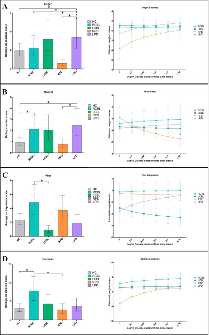
There is growing evidence that both the basal ganglia and the cerebellum play functional roles in emotion processing, either directly or indirectly, through their connections with cortical and subcortical structures. However, the lateralization of this complex processing in emotion recognition remains unclear. To address this issue, we investigated emotional prosody recognition in individuals with Parkinson's disease (model of basal ganglia dysfunction) or cerebellar stroke patients, as well as in matched healthy controls (n = 24 in each group). We analysed performances according to the lateralization of the predominant brain degeneration/lesion. Results showed that a right (basal ganglia and cerebellar) hemispheric dysfunction was likely to induce greater deficits than a left one. Moreover, deficits following left hemispheric dysfunction were only observed in cerebellar stroke patients, and these deficits resembled those observed after degeneration of the right basal ganglia. Additional analyses taking disease duration / time since stroke into consideration revealed a worsening of performances in patients with predominantly right-sided lesions over time. These results point to the differential, but complementary, involvement of the cerebellum and basal ganglia in emotional prosody decoding, with a probable hemispheric specialization according to the level of cognitive integration.
越来越多的证据表明,基底神经节和小脑在情绪加工中发挥着功能作用,它们通过与皮质和皮质下结构的连接,直接或间接地参与其中。然而,这种复杂的情绪识别加工过程的脑区偏侧化仍不清楚。为了解决这个问题,我们研究了帕金森病患者(基底神经节功能障碍模型)、小脑中风患者以及匹配的健康对照者(每组n = 24)的情绪韵律识别能力。我们根据主要脑区变性/损伤的偏侧化情况分析了他们的表现。结果表明,右侧(基底神经节和小脑)半球功能障碍可能比左侧半球功能障碍导致更严重的缺陷。此外,仅在小脑中风患者中观察到左侧半球功能障碍后的缺陷,这些缺陷与右侧基底神经节变性后观察到的缺陷相似。考虑到疾病持续时间/中风后的时间进行的进一步分析显示,主要为右侧病变的患者的表现会随着时间的推移而恶化。这些结果表明,小脑和基底神经节在情绪韵律解码中存在差异但互补的参与,并且根据认知整合水平可能存在半球特化。