Al-Kadi Mohammad, AlOtieschan Salman, Almahdi Mohammad Jihad, AlHajress Rafeef
Division of Otolaryngology - Head & Neck Surgery, Department of Surgery, King Abdulaziz Medical City, Riyadh, SAU.
Medicine, College of Medicine, King Saud Bin Abdulaziz University for Health Sciences, Riyadh, SAU.
Cureus. 2022 Mar 21;14(3):e23348. doi: 10.7759/cureus.23348. eCollection 2022 Mar.
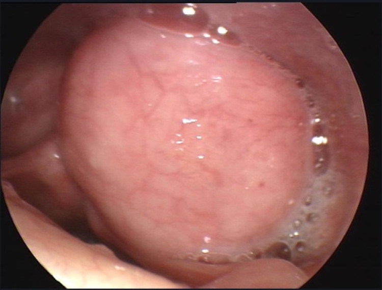
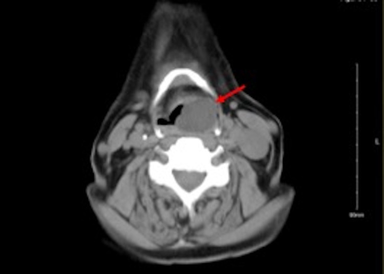
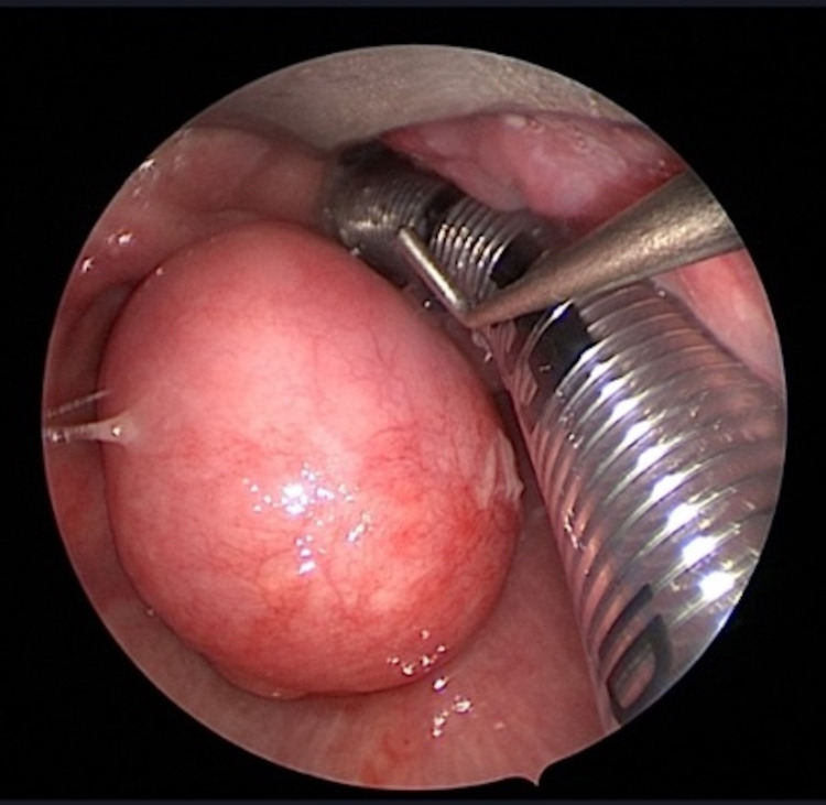
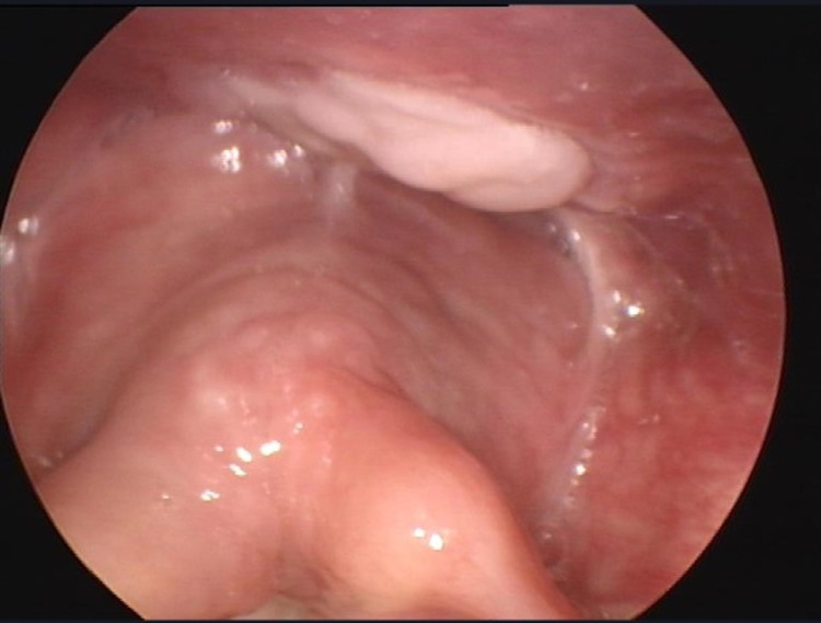
Nearly 20% of all mesenchymal tumors are liposarcoma in origin, mostly occurring in extremities and trunk. However, few cases of liposarcoma in the hypopharynx have been documented. This atypical localization of liposarcoma warrants a great interest in reporting such a case. Here, we report an 81-year-old Saudi male who presented to our clinic complaining of progressive dysphagia and globus sensation for two months. On examination, using a flexible nasopharyngoscopy, a hypopharyngeal mass occupying the left piriform sinus originating from the mucosa of the posterior pharyngeal wall and anteriorly from the anterior and medial piriform sinus mucosa was observed. Contrasted head and neck CT-scan revealed a benign-looking well-defined left-sided submucosal cyst aligned along the left aryepiglottic fold encroaching and narrowing the laryngeal inlet with dimensions of 1.8×2.1×2.7 cm. The mass was resected successfully using a trans-oral approach. A histopathological review showed spindle stromal cells that reacted positively for CD34 (Qbend10) on immunohistochemical staining and positive result for MDM2 (12q15) Amp. The pathology result indicates an abnormal amplification of the MDM2 gene region. The patient was followed for almost two years without evidence of recurrence. In conclusion, atypical lipomatous tumors (ALTs) of the hypopharynx are rarely diagnosed, and the gold standard for diagnosis is biopsy. Transoral endoscopic approach has a better outcome than cervical approach. Follow-up of patients with ALT is crucial, due to the highly recurring nature of the disease. Here we present a rare case of ALT, the patient had complete remission without complication.
所有间充质肿瘤中近20%起源于脂肪肉瘤,主要发生在四肢和躯干。然而,下咽脂肪肉瘤的病例报道很少。脂肪肉瘤的这种非典型定位使得报道这样一个病例备受关注。在此,我们报告一名81岁的沙特男性,他因进行性吞咽困难和咽部异物感两个月前来我院就诊。检查时,通过软性鼻咽镜观察到一个占据左侧梨状窦的下咽肿物,其起源于咽后壁黏膜,前部来自梨状窦前内侧黏膜。头颈增强CT扫描显示一个边界清晰、外观良性的左侧黏膜下囊肿,沿左杓会厌襞排列,侵犯并狭窄喉入口,大小为1.8×2.1×2.7 cm。采用经口入路成功切除肿物。组织病理学检查显示梭形间质细胞,免疫组化染色CD34(Qbend10)呈阳性,MDM2(12q15)扩增呈阳性。病理结果表明MDM2基因区域存在异常扩增。对该患者进行了近两年的随访,无复发迹象。总之,下咽非典型脂肪瘤性肿瘤(ALT)很少被诊断出来,诊断的金标准是活检。经口内镜入路比颈部入路效果更好。由于该疾病具有高度复发性,对ALT患者进行随访至关重要。在此我们报告一例罕见的ALT病例,患者完全缓解且无并发症。