Yang Zhengshi, Caldwell Jessica Z K, Cummings Jeffrey L, Ritter Aaron, Kinney Jefferson W, Cordes Dietmar
Cleveland Clinic Lou Ruvo Center for Brain Health, Las Vegas, NV, United States.
Department of Brain Health, University of Nevada Las Vegas, Las Vegas, NV, United States.
Front Psychiatry. 2022 Apr 5;13:804168. doi: 10.3389/fpsyt.2022.804168. eCollection 2022.
To assess the pathological aging effect on caudate functional connectivity among mild cognitive impairment (MCI) participants and examine whether and how sex and amyloid contribute to this process.
Two hundred and seventy-seven functional magnetic resonance imaging (fMRI) sessions from 163 cognitive normal (CN) older adults and 309 sessions from 139 participants with MCI were included as the main sample in our analysis. Pearson's correlation was used to characterize the functional connectivity (FC) between caudate nuclei and each brain region, then caudate nodal strength was computed to quantify the overall caudate FC strength. Association analysis between caudate nodal strength and age was carried out in MCI and CN separately using linear mixed effect (LME) model with covariates (education, handedness, sex, Apolipoprotein E4, and intra-subject effect). Analysis of covariance was conducted to investigate sex, amyloid status, and their interaction effects on aging with the fMRI data subset having amyloid status available. LME model was applied to women and men separately within MCI group to evaluate aging effects on caudate nodal strength and each region's connectivity with caudate nuclei. We then evaluated the roles of sex and amyloid status in the associations of neuropsychological scores with age or caudate nodal strength. An independent cohort was used to validate the sex-dependent aging effects in MCI.
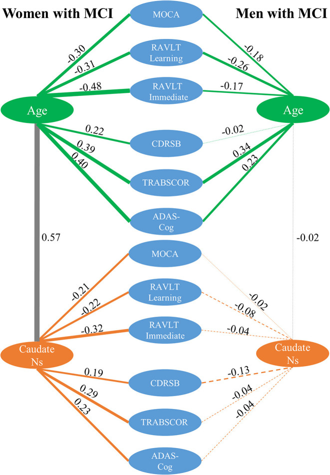
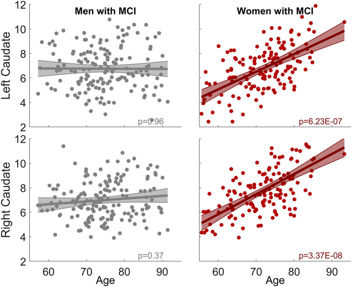
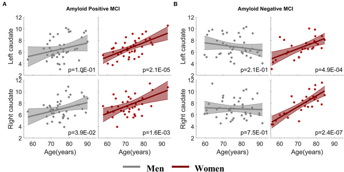
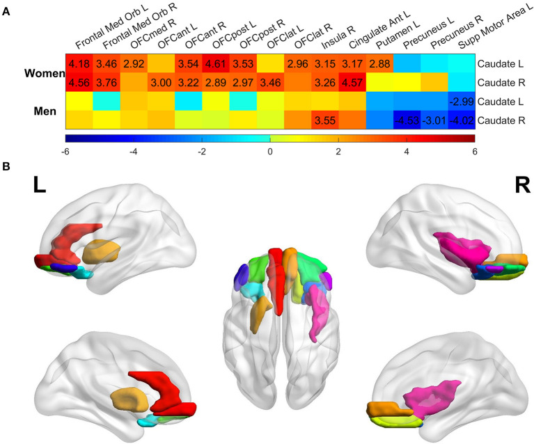
The MCI group had significantly stronger age-related increase of caudate nodal strength compared to the CN group. Analyzing women and men separately revealed that the aging effect on caudate nodal strength among MCI participants was significant only for women (left: = 6.23 × 10, right: = 3.37 × 10), but not for men ( > 0.3 for bilateral caudate nuclei). The aging effects on caudate nodal strength were not significantly mediated by brain amyloid burden. Caudate connectivity with ventral prefrontal cortex substantially contributed to the aging effect on caudate nodal strength in women with MCI. Higher caudate nodal strength is significantly related to worse cognitive performance in women but not in men with MCI.
Sex modulates the pathological aging effects on caudate nodal strength in MCI regardless of amyloid status. Caudate nodal strength may be a sensitive biomarker of pathological aging in women with MCI.
评估轻度认知障碍(MCI)参与者尾状核功能连接的病理衰老效应,并研究性别和淀粉样蛋白是否以及如何影响这一过程。
我们的分析主要样本包括163名认知正常(CN)老年人的277次功能磁共振成像(fMRI)扫描,以及139名MCI参与者的309次扫描。使用Pearson相关性分析来表征尾状核与每个脑区之间的功能连接(FC),然后计算尾状核节点强度以量化整体尾状核FC强度。在MCI和CN组中分别使用线性混合效应(LME)模型(包括协变量:教育程度、利手、性别、载脂蛋白E4和个体内效应)对尾状核节点强度与年龄进行关联分析。对于有淀粉样蛋白状态数据的fMRI数据子集,进行协方差分析以研究性别、淀粉样蛋白状态及其对衰老的交互作用。在MCI组内分别对女性和男性应用LME模型,以评估衰老对尾状核节点强度以及每个脑区与尾状核连接的影响。然后,我们评估了性别和淀粉样蛋白状态在神经心理学评分与年龄或尾状核节点强度关联中的作用。使用一个独立队列验证MCI中性别依赖性衰老效应。
与CN组相比,MCI组尾状核节点强度的年龄相关增加显著更强。分别对女性和男性进行分析发现,MCI参与者中衰老对尾状核节点强度的影响仅在女性中显著(左侧: = 6.23×10,右侧: = 3.37×10),而在男性中不显著(双侧尾状核 > 0.3)。脑淀粉样蛋白负担并未显著介导衰老对尾状核节点强度的影响。在患有MCI的女性中,尾状核与腹侧前额叶皮层的连接对衰老对尾状核节点强度的影响有显著贡献。在患有MCI的女性中,较高的尾状核节点强度与较差的认知表现显著相关,而在男性中则不然。
无论淀粉样蛋白状态如何,性别都会调节MCI中尾状核节点强度的病理衰老效应。尾状核节点强度可能是患有MCI的女性病理衰老的敏感生物标志物。