School of Biological Sciences, Nanyang Technological University, 60 Nanyang Drive, Singapore 637551, Singapore.
NTU Institute of Structural Biology, Nanyang Technological University, 59 Nanyang Drive, Singapore 636921, Singapore.
Nucleic Acids Res. 2022 May 20;50(9):5047-5063. doi: 10.1093/nar/gkac309.
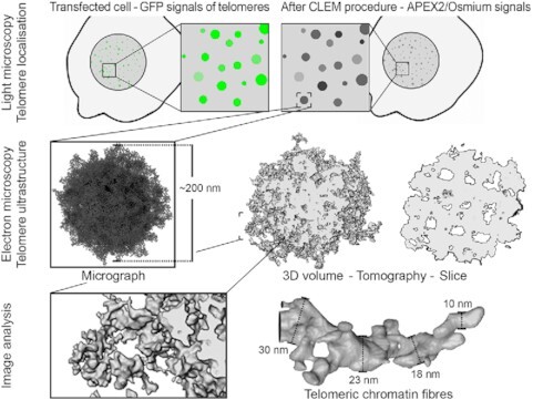
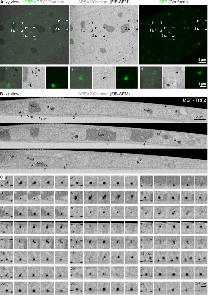
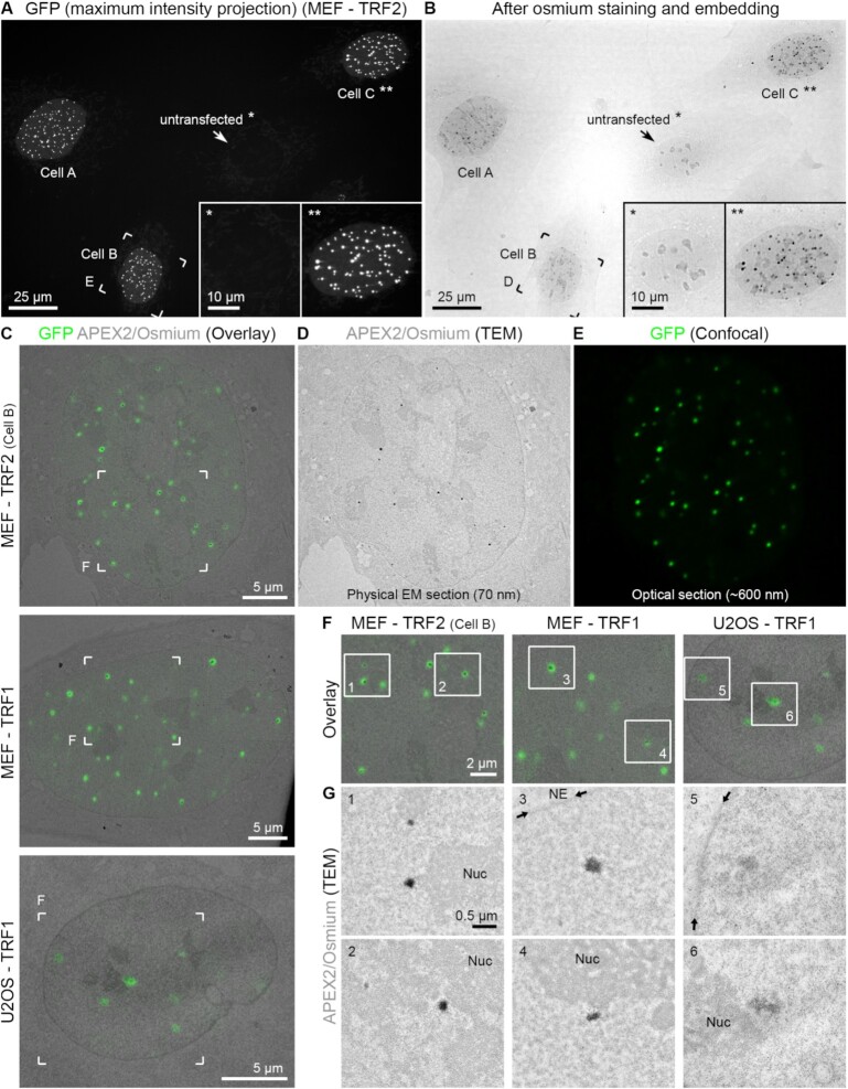
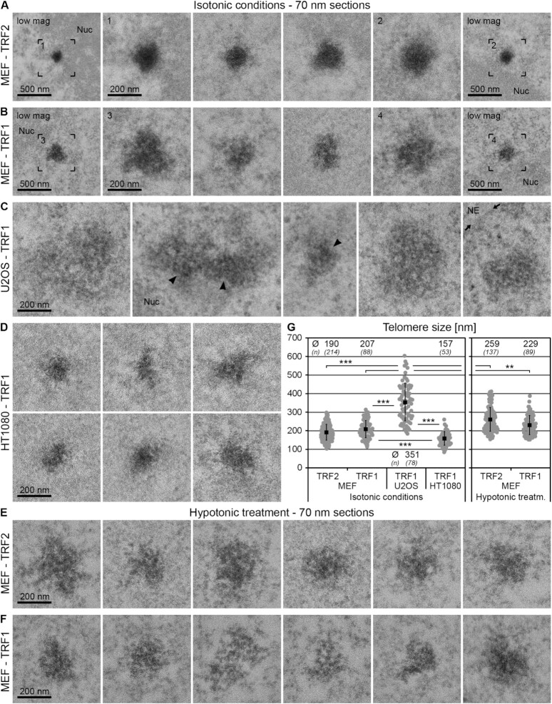
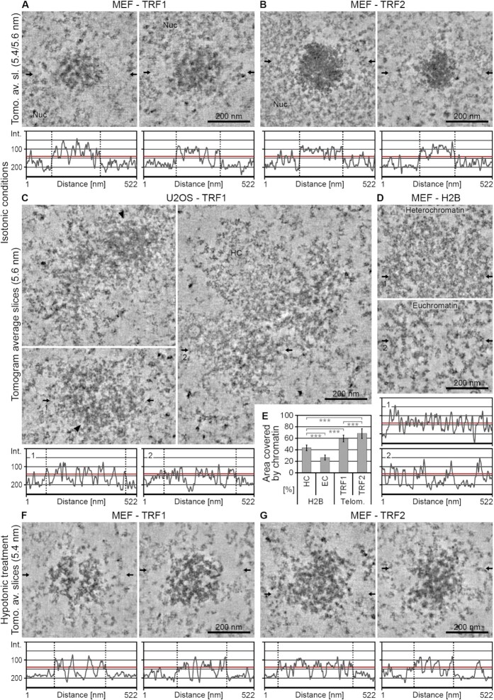
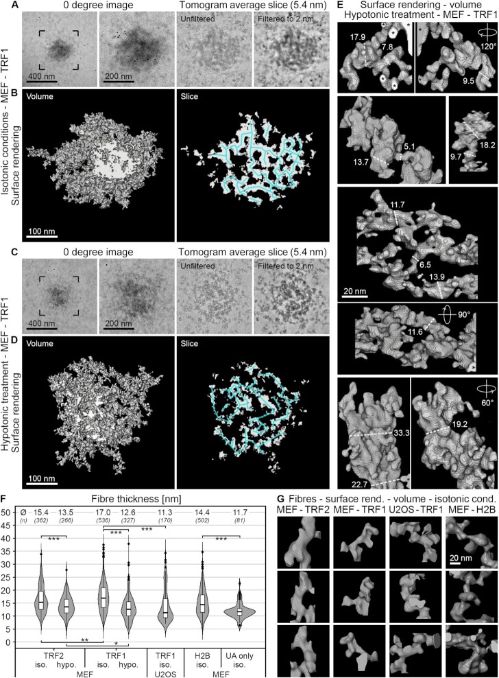
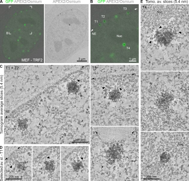
Telomeres, the ends of linear chromosomes, are composed of repetitive DNA sequences, histones and a protein complex called shelterin. How DNA is packaged at telomeres is an outstanding question in the field with significant implications for human health and disease. Here, we studied the architecture of telomeres and their spatial association with other chromatin domains in different cell types using correlative light and electron microscopy. To this end, the shelterin protein TRF1 or TRF2 was fused in tandem to eGFP and the peroxidase APEX2, which provided a selective and electron-dense label to interrogate telomere organization by transmission electron microscopy, electron tomography and scanning electron microscopy. Together, our work reveals, for the first time, ultrastructural insight into telomere architecture. We show that telomeres are composed of a dense and highly compacted mesh of chromatin fibres. In addition, we identify marked differences in telomere size, shape and chromatin compaction between cancer and non-cancer cells and show that telomeres are in direct contact with other heterochromatin regions. Our work resolves the internal architecture of telomeres with unprecedented resolution and advances our understanding of how telomeres are organized in situ.
端粒是线性染色体的末端,由重复 DNA 序列、组蛋白和一种称为 shelterin 的蛋白质复合物组成。端粒处的 DNA 如何包装是该领域的一个悬而未决的问题,对人类健康和疾病有重大影响。在这里,我们使用相关的光和电子显微镜研究了不同细胞类型中端粒的结构及其与其他染色质域的空间关联。为此,将 shelterin 蛋白 TRF1 或 TRF2 串联融合到 eGFP 和过氧化物酶 APEX2 上,这为通过透射电子显微镜、电子断层扫描和扫描电子显微镜研究端粒组织提供了选择性和电子致密标记。总之,我们的工作首次揭示了端粒结构的超微结构见解。我们表明,端粒由密集且高度紧凑的染色质纤维网格组成。此外,我们还发现了癌症和非癌细胞中端粒大小、形状和染色质紧缩的显著差异,并表明端粒与其他异染色质区域直接接触。我们的工作以空前的分辨率解决了端粒的内部结构,并推进了我们对端粒在原位组织方式的理解。