Department of Translational Molecular Pathology, University of Texas MD Anderson Cancer Center, Houston, Texas.
Department of Biostatistics, University of Texas MD Anderson Cancer Center, Houston, Texas.
Clin Cancer Res. 2022 May 2;28(9):1938-1947. doi: 10.1158/1078-0432.CCR-21-2585.
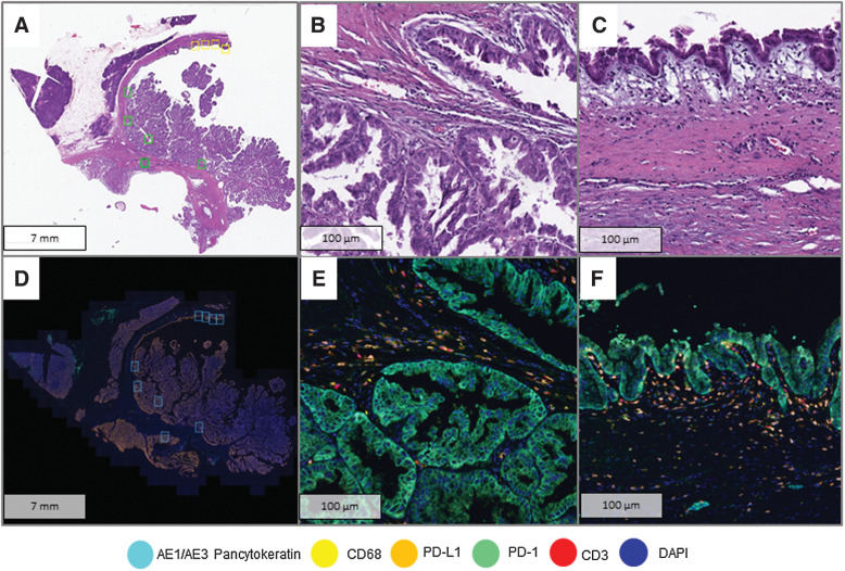
Intraductal papillary mucinous neoplasms (IPMN) are bona fide precursors to pancreatic ductal adenocarcinoma (PDAC). While genomic alterations during multistep IPMN progression have been well cataloged, the accompanying changes within the tumor immune microenvironment (TIME) have not been comprehensively studied. Herein, we investigated TIME-related alterations during IPMN progression, using multiplex immunofluorescence (mIF) coupled with high-resolution image analyses.
Two sets of formalin-fixed, paraffin-embedded tissue samples from surgically resected IPMNs were analyzed. The training set of 30 samples consisted of 11 low-grade IPMN (LG-IPMN), 17 high-grade IPMN (HG-IPMN), and 2 IPMN with PDAC, while a validation set of 93 samples comprised of 55 LG-IPMN and 38 HG-IPMN. The training set was analyzed with two panels of immuno-oncology-related biomarkers, while the validation set was analyzed with a subset of markers found significantly altered in the training set.
Cell types indicative of enhanced immune surveillance, including cytotoxic and memory T cells, and antigen-experienced T cells and B cells, were all found at higher densities within isolated LG-IPMNs compared with HG-IPMNs. Notably, the TIME of LG-IPMNs that had progressed at the time of surgical resection (progressor LGD) resembled that of the synchronous HG-IPMNs, underscoring that attenuated immune surveillance occurs even in LG-IPMNs destined for progression.
Our findings provide a basis for interception of cystic neoplasia to PDAC, through maintenance of sustained immune surveillance using vaccines and other prevention approaches.
导管内乳头状黏液性肿瘤(IPMN)是胰腺导管腺癌(PDAC)的真正前体。虽然多步骤 IPMN 进展过程中的基因组改变已得到很好的编目,但肿瘤免疫微环境(TIME)中的伴随变化尚未得到全面研究。在此,我们使用多重免疫荧光(mIF)结合高分辨率图像分析,研究了 IPMN 进展过程中的 TIME 相关改变。
分析了来自手术切除的 IPMN 的两组福尔马林固定、石蜡包埋的组织样本。包含 30 个样本的训练集包括 11 个低级别 IPMN(LG-IPMN)、17 个高级别 IPMN(HG-IPMN)和 2 个伴有 PDAC 的 IPMN,而包含 93 个样本的验证集包括 55 个 LG-IPMN 和 38 个 HG-IPMN。训练集使用两个免疫肿瘤学相关生物标志物面板进行分析,而验证集则使用在训练集中发现明显改变的一组标志物进行分析。
在分离的 LG-IPMN 中,包括细胞毒性和记忆 T 细胞以及抗原经验的 T 细胞和 B 细胞在内的提示增强免疫监视的细胞类型的密度均高于 HG-IPMN。值得注意的是,在手术切除时已经进展的 LG-IPMN 的 TIME(进展 LGD)与同步 HG-IPMN 的 TIME 相似,这表明即使在注定要进展的 LG-IPMN 中,免疫监视也会减弱。
我们的研究结果为通过使用疫苗和其他预防方法维持持续的免疫监视,为阻断囊性肿瘤向 PDAC 的进展提供了基础。