Bal Christina, Göschl Lisa, Milos Ruxandra-Iulia, Gerstbrein Klaus, Kerschbaumer Andreas, Idzko Marco, Gompelmann Daniela
Department of Medicine II, Division of Pulmonology, Medical University of Vienna, Vienna, Austria.
Department of Medicine III, Division of Rheumatology, Medical University of Vienna, Vienna, Austria.
Respir Med Case Rep. 2022 Apr 16;37:101650. doi: 10.1016/j.rmcr.2022.101650. eCollection 2022.
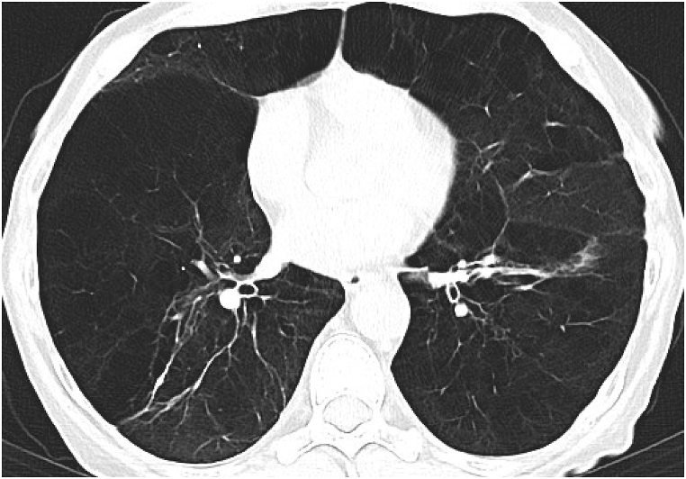
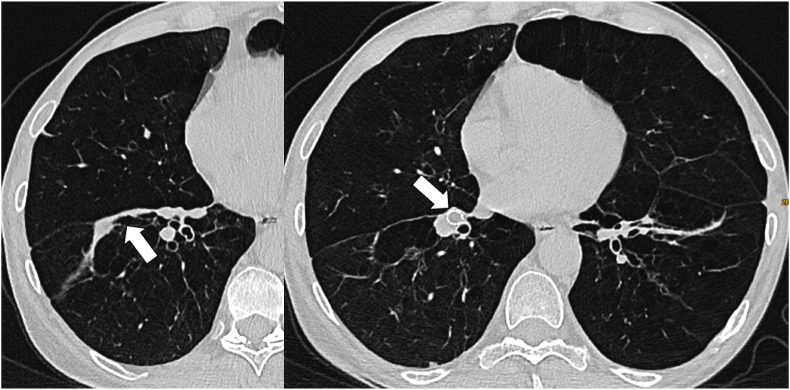
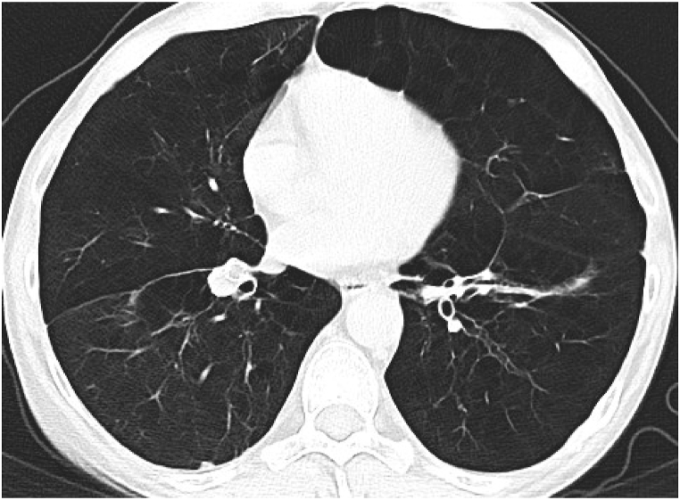
A 53-year old female patient with history of hypocomplementaemic urticarial vasculitis syndrome (HUVS) and polyarteritis nodosa presented with progressive dyspnoea on exertion due to emphysema. Lung function revealed a severe obstructive ventilator disorder with a forced expiratory volume in 1 second of 22% of predicted, and a significant hyperinflation with a residual volume of 321% of predicted. Multi-detector computed tomography (MDCT) scan and quantitative CT analysis (StratX software) confirmed a lower lobe predominant emphysema. Considering the young age, the very severely impaired lung function, the relatively low nicotine abuse, the exclusion of alpha-1 antitrypsin deficiency, together with the known diagnosis of HUVS, the emphysema was more likely due to the vasculitis than to a typical chronic obstructive lung disease. MDCT scan showed that particularly the segment 8 of the right lower lobe was severely emphysematous destroyed and hyperinflated. Invasive Chartis® measurement revealed no significant collateral ventilation of the isolated segment 8 of the right lower lobe, so that an endobronchial valve placement was performed. Three months following intervention, the MDCT scan revealed a complete collapse of the segment 8 on the right, which was associated with a significant clinical benefit and a mild reduction of the hyperinflation in the lung function test.
一名53岁女性患者,有低补体血症性荨麻疹性血管炎综合征(HUVS)和结节性多动脉炎病史,因肺气肿出现进行性劳力性呼吸困难。肺功能显示严重的阻塞性通气障碍,一秒用力呼气量为预测值的22%,且有明显的肺过度充气,残气量为预测值的321%。多排螺旋计算机断层扫描(MDCT)和定量CT分析(StratX软件)证实肺气肿以两下肺为主。考虑到患者年轻、肺功能严重受损、尼古丁滥用程度相对较低、排除α-1抗胰蛋白酶缺乏,以及已知的HUVS诊断,肺气肿更可能是由血管炎引起,而非典型的慢性阻塞性肺疾病。MDCT扫描显示,尤其是右下叶8段严重肺气肿性破坏且肺过度充气。侵入性Chartis®测量显示右下叶孤立的8段无明显的侧支通气,因此进行了支气管内瓣膜置入。干预三个月后,MDCT扫描显示右侧8段完全萎陷,这与显著的临床获益以及肺功能测试中肺过度充气的轻度减轻相关。