Pina Violeta, Campello Víctor M, Lekadir Karim, Seguí Santi, García-Santos Jose M, Fuentes Luis J
Departamento de Psicología Evolutiva y de la Educación, Facultad de Educación, Economía y Tecnología de Ceuta, Universidad de Granada, Ceuta, Spain.
Departament de Matemàtiques i Informàtica, Universitat de Barcelona, Barcelona, Spain.
Front Neurosci. 2022 Apr 14;16:819069. doi: 10.3389/fnins.2022.819069. eCollection 2022.
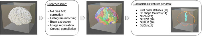
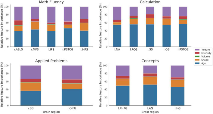
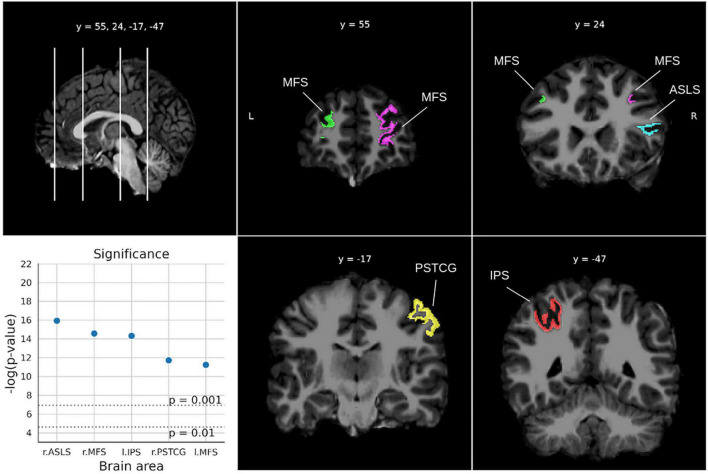
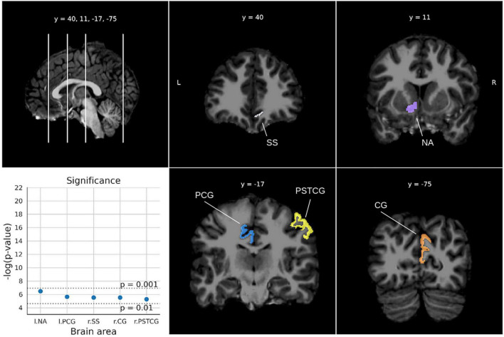
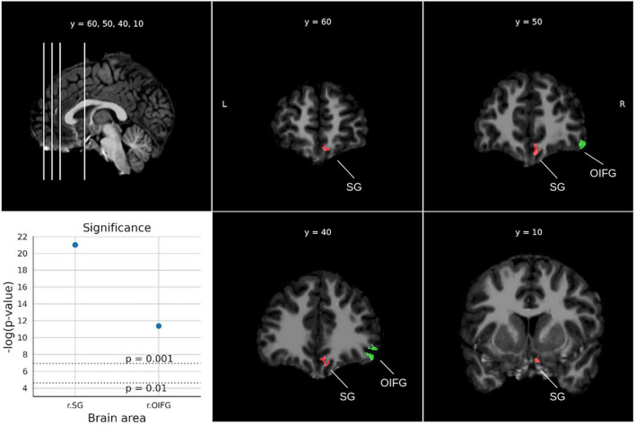
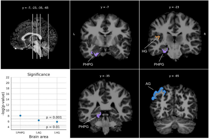
Structural magnetic resonance imaging (sMRI) studies have shown that children that differ in some mathematical abilities show differences in gray matter volume mainly in parietal and frontal regions that are involved in number processing, attentional control, and memory. In the present study, a structural neuroimaging analysis based on radiomics and machine learning models is presented with the aim of identifying the brain areas that better predict children's performance in a variety of mathematical tests. A sample of 77 school-aged children from third to sixth grade were administered four mathematical tests: Math fluency, Calculation, Applied problems and Quantitative concepts as well as a structural brain imaging scan. By extracting radiomics related to the shape, intensity, and texture of specific brain areas, we observed that areas from the frontal, parietal, temporal, and occipital lobes, basal ganglia, and limbic system, were differentially related to children's performance in the mathematical tests. sMRI-based analyses in the context of mathematical performance have been mainly focused on volumetric measures. However, the results for radiomics-based analysis showed that for these areas, texture features were the most important for the regression models, while volume accounted for less than 15% of the shape importance. These findings highlight the potential of radiomics for more in-depth analysis of medical images for the identification of brain areas related to mathematical abilities.
结构磁共振成像(sMRI)研究表明,在某些数学能力上存在差异的儿童,其灰质体积的差异主要体现在顶叶和额叶区域,这些区域参与数字处理、注意力控制和记忆。在本研究中,我们提出了一种基于放射组学和机器学习模型的结构神经影像学分析方法,旨在识别能更好预测儿童在各种数学测试中表现的脑区。我们对77名三至六年级的学龄儿童进行了四项数学测试:数学流畅性、计算、应用题和定量概念测试,并对他们进行了脑部结构成像扫描。通过提取与特定脑区的形状、强度和纹理相关的放射组学特征,我们观察到额叶、顶叶、颞叶、枕叶、基底神经节和边缘系统的区域与儿童在数学测试中的表现存在不同程度的关联。在数学表现的背景下,基于sMRI的分析主要集中在体积测量上。然而,基于放射组学的分析结果表明,对于这些区域,纹理特征对回归模型最为重要,而体积在形状重要性方面所占比例不到15%。这些发现凸显了放射组学在更深入分析医学图像以识别与数学能力相关脑区方面的潜力。