Zhu Qianqian, Tang Shengluan, Zhu Yanwen, Chen Di, Huang Jialyu, Lin Jiaying
Department of Assisted Reproduction, Shanghai Ninth People's Hospital, Shanghai Jiao Tong University School of Medicine, Shanghai, China.
Center for Reproductive Medicine, Jiangxi Maternal and Child Health Hospital, Nanchang University School of Medicine, Nanchang, China.
Front Bioeng Biotechnol. 2022 Apr 13;10:868734. doi: 10.3389/fbioe.2022.868734. eCollection 2022.
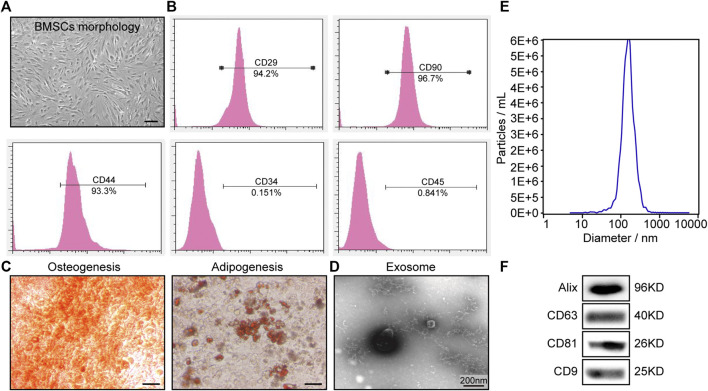
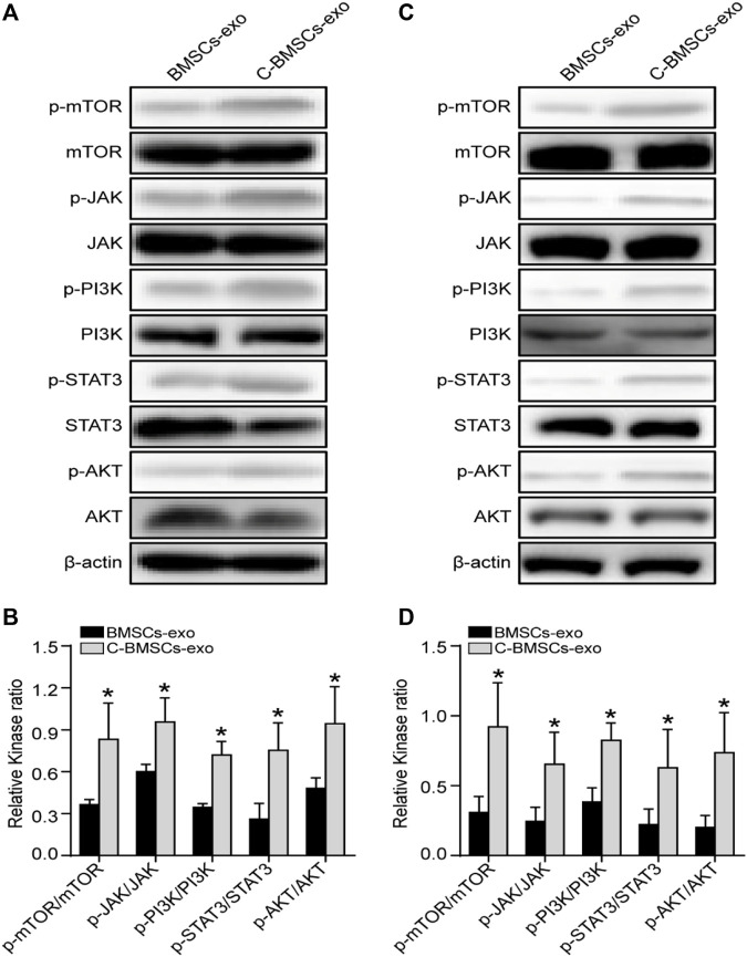
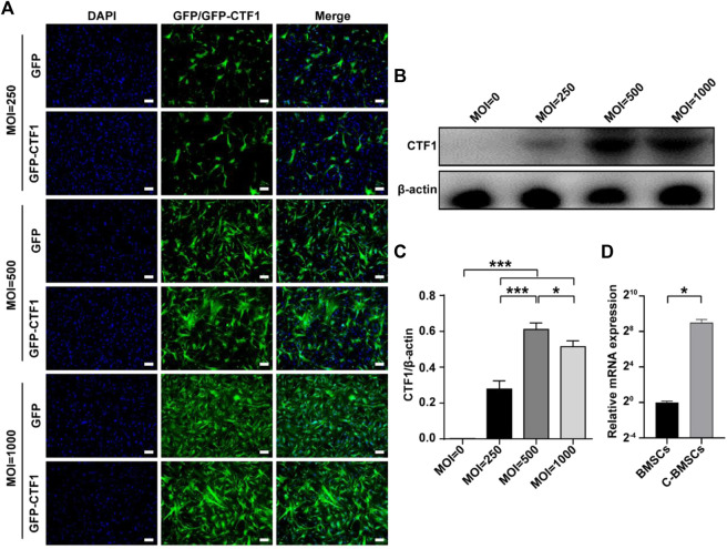
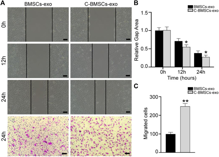
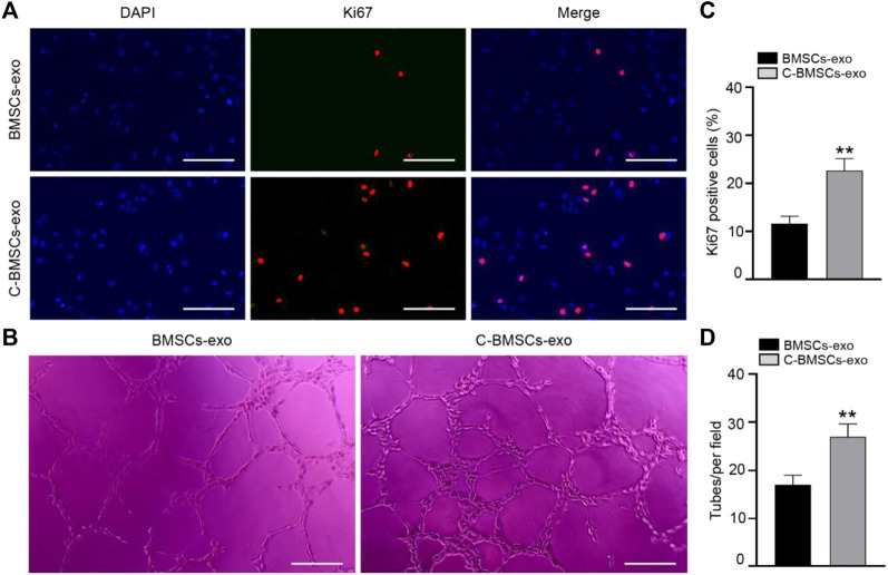
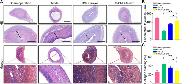
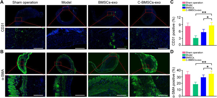
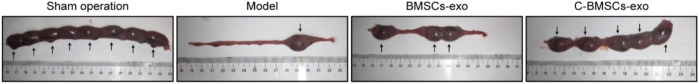
Thin endometrial tissue is a leading cause of embryo transfer failure, potentially contributing to sustained infertility and associated adverse outcomes. The application of exosomes derived from autologous or allogeneic bone marrow-derived stem cells (BMSCs) has been used to promote uterine repair following injury, and there is also prior evidence that stem cell transplantation can bolster fertility. Genetic modifications represent a primary approach to enhancing exosomal therapy strategies. The present study thus explored the effects of Cardiotrophin-1 (CTF1)-modified BMSCs-exo on fertility-related outcomes. An adenoviral vector was used to generate CTF1-overexpressing BMSCs (C-BMSCs), after which exosomes were isolated from control BMSCs (BMSC-exos) and C-BMSCs (C-BMSC-exos). The angiogenic effects of C-BMSC-exo treatment were assessed through analyses of endothelial cell proliferation and tube formation. Model rats exhibiting endometrial thinning were administered C-BMSCs-exo, after which the effects of such treatment were assessed through H&E staining, Masson's trichrome staining, and immunofluorescence analyses. The mechanistic basis for the proangiogenic effects of CTF1 as a driver of endometrial regeneration was additionally explored. C-BMSC-exo treatment of HUVECs was associated with enhanced neovascularization, as evidenced by improved proliferation, migration, and tube formation. Importantly, such treatment was also linked to tissue regeneration, neovascularization, and the suppression of localized tissue fibrosis . Regenerated endometrial tissue exhibited higher embryo receptivity and was associated with higher birth rates in treated rats. The upregulation of the JAK/PI3K/mTOR/STAT3 signaling pathways in C-BMSC-exo-treated rats may underscore the mechanistic basis whereby CTF1 can positively impact endometrial angiogenesis and regeneration. Our data suggest that exosomes produced by CTF1-modified BMSCs can more effectively promote the regeneration of endometrial and myometrial tissues, driving neovascularization in a manner that improves endometrial receptivity in a rat model system, highlighting the therapeutic promise of this approach for patients diagnosed with endometrial thinning.
薄型子宫内膜组织是胚胎移植失败的主要原因,可能导致持续性不孕及相关不良后果。应用自体或异体骨髓来源干细胞(BMSC)分泌的外泌体已被用于促进损伤后的子宫修复,并且先前也有证据表明干细胞移植可提高生育能力。基因修饰是增强外泌体治疗策略的主要方法。因此,本研究探讨了心肌营养素-1(CTF1)修饰的BMSC来源外泌体(C-BMSC-exo)对生育相关结局的影响。使用腺病毒载体生成过表达CTF1的BMSC(C-BMSC),之后从对照BMSC(BMSC-exo)和C-BMSC(C-BMSC-exo)中分离出外泌体。通过分析内皮细胞增殖和管腔形成来评估C-BMSC-exo处理的血管生成作用。对表现出子宫内膜变薄的模型大鼠给予C-BMSC-exo,之后通过苏木精-伊红染色、Masson三色染色和免疫荧光分析来评估该处理的效果。还额外探讨了CTF1作为子宫内膜再生驱动因子发挥促血管生成作用的机制基础。C-BMSC-exo处理人脐静脉内皮细胞(HUVEC)与新生血管形成增强相关,表现为增殖、迁移和管腔形成改善。重要的是,这种处理还与组织再生、新生血管形成以及局部组织纤维化的抑制有关。再生的子宫内膜组织表现出更高的胚胎接受性,并且与处理后大鼠的更高出生率相关。C-BMSC-exo处理的大鼠中JAK/PI3K/mTOR/STAT3信号通路的上调可能强调了CTF1能够积极影响子宫内膜血管生成和再生的机制基础。我们的数据表明,CTF1修饰的BMSC产生的外泌体能够更有效地促进子宫内膜和肌层组织的再生,以改善大鼠模型系统中子宫内膜接受性的方式驱动新生血管形成,突出了该方法对诊断为子宫内膜变薄患者的治疗前景。