• 文献检索
  • 文档翻译
  • 深度研究
  • 学术资讯
  • Suppr Zotero 插件Zotero 插件
  • 邀请有礼
  • 套餐&价格
  • 历史记录
应用&插件
Suppr Zotero 插件Zotero 插件浏览器插件Mac 客户端Windows 客户端微信小程序
定价
高级版会员购买积分包购买API积分包
服务
文献检索文档翻译深度研究API 文档MCP 服务
关于我们
关于 Suppr公司介绍联系我们用户协议隐私条款
关注我们

Suppr 超能文献

核心技术专利:CN118964589B侵权必究
粤ICP备2023148730 号-1Suppr @ 2026

文献检索

告别复杂PubMed语法,用中文像聊天一样搜索,搜遍4000万医学文献。AI智能推荐,让科研检索更轻松。

立即免费搜索

文件翻译

保留排版,准确专业,支持PDF/Word/PPT等文件格式,支持 12+语言互译。

免费翻译文档

深度研究

AI帮你快速写综述,25分钟生成高质量综述,智能提取关键信息,辅助科研写作。

立即免费体验

近视眼中视网膜中央动脉的血流动力学和形态学变化。

Hemodynamic and morphological changes of the central retinal artery in myopic eyes.

机构信息

School of Optometry, Faculty of Health and Social Science, The Hong Kong Polytechnic University, Hung Hom, Kowloon, Hong Kong, China.

Research Centre for SHARP Vision, The Hong Kong Polytechnic University, Hung Hom, Kowloon, Hong Kong, China.

出版信息

Sci Rep. 2022 May 2;12(1):7104. doi: 10.1038/s41598-022-11087-x.

DOI:10.1038/s41598-022-11087-x
PMID:35501327
原文链接:https://pmc.ncbi.nlm.nih.gov/articles/PMC9061854/
Abstract

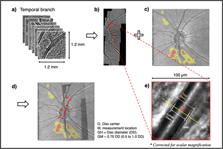
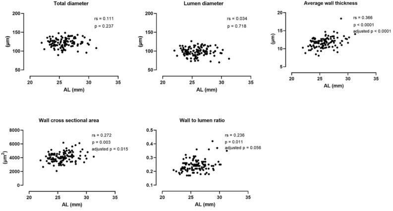
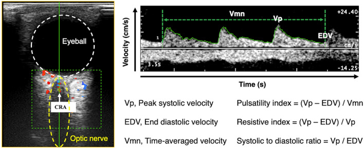
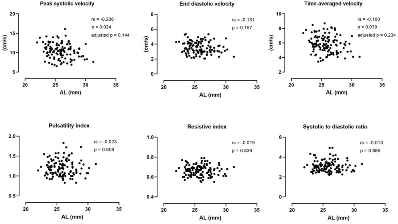
Due to excessive elongation of the eyeball, myopia-related vascular abnormalities are frequently observed in the central retinal artery (CRA) and its intraretinal branches. In addition to inconsistency in previously reported findings, hemodynamic (reduced flow velocity, increased vascular resistance) and morphological changes (narrower vessel diameter) were usually studied separately. This cross-sectional study evaluated the hemodynamic and morphological characteristics concurrently in a large sample of healthy myopes, by using the color Doppler ultrasound and adaptive optics retinal camera. Results showed that the retrobulbar segment of CRA had a tendency of slightly reduced flow velocity in eyeballs with longer axial length, but the correlation was not significant after adjusting for the multiple correlations. Vascular resistance was not affected by the axial elongation. With respect to the intraretinal branches, no significant changes in longer eyes of total diameter or lumen diameter were observed, while both the wall thickness and the wall cross-sectional area were significantly increased, but only a marginally increase in the wall to lumen ratio was found with increasing axial length. This implies some potential small artery remodeling in the intraretinal CRA branches. Overall, blood supply of the inner retina in healthy young myopes is likely to be maintained. Additionally, morphological parameters of vascular microstructure could be potential biomarkers to monitor myopia progression and understand myopia-related vascular abnormalities in future studies.

摘要

由于眼球过度伸长,近视相关的血管异常经常在视网膜中央动脉(CRA)及其视网膜内分支中观察到。除了先前报道的结果不一致外,血流动力学(血流速度降低,血管阻力增加)和形态学变化(血管直径变窄)通常是分开研究的。这项横断面研究通过彩色多谱勒超声和自适应光学视网膜照相机,在大量健康近视者中同时评估了血流动力学和形态特征。结果表明,在眼轴较长的眼球中,CRA 的球后段血流速度有略微降低的趋势,但在调整多重相关性后,相关性并不显著。血管阻力不受轴向伸长的影响。对于视网膜内分支,在更长的总直径或管腔直径的眼睛中没有观察到明显的变化,而壁厚度和壁横截面积显著增加,但是随着轴向长度的增加,仅发现壁腔比略有增加。这意味着视网膜内 CRA 分支中可能存在一些潜在的小动脉重塑。总的来说,健康年轻近视者的内视网膜血液供应可能得到维持。此外,血管微观结构的形态参数可能是未来研究中监测近视进展和了解近视相关血管异常的潜在生物标志物。

https://cdn.ncbi.nlm.nih.gov/pmc/blobs/fc54/9061854/1ae54dd3c23b/41598_2022_11087_Fig4_HTML.jpg
https://cdn.ncbi.nlm.nih.gov/pmc/blobs/fc54/9061854/704252c76b34/41598_2022_11087_Fig1_HTML.jpg
https://cdn.ncbi.nlm.nih.gov/pmc/blobs/fc54/9061854/50aafd3ef7bf/41598_2022_11087_Fig2_HTML.jpg
https://cdn.ncbi.nlm.nih.gov/pmc/blobs/fc54/9061854/09622f1aa5f5/41598_2022_11087_Fig3_HTML.jpg
https://cdn.ncbi.nlm.nih.gov/pmc/blobs/fc54/9061854/1ae54dd3c23b/41598_2022_11087_Fig4_HTML.jpg

相似文献

1
Hemodynamic and morphological changes of the central retinal artery in myopic eyes.近视眼中视网膜中央动脉的血流动力学和形态学变化。
Sci Rep. 2022 May 2;12(1):7104. doi: 10.1038/s41598-022-11087-x.
2
[Evaluation of the arterial blood flow parameters in the eye of myopic patients].[近视患者眼部动脉血流参数的评估]
Pol Merkur Lekarski. 2013 Apr;34(202):205-9.
3
Ocular blood flow measurements in healthy human myopic eyes.健康人近视眼的眼血流测量。
Graefes Arch Clin Exp Ophthalmol. 2010 Nov;248(11):1587-94. doi: 10.1007/s00417-010-1407-9. Epub 2010 May 26.
4
Correlation between retrobulbar blood flow parameters and retinal nerve fiber, ganglion cell and inner plexus layer thickness in myopia.近视患者球后血流参数与视网膜神经纤维、节细胞和内丛状层厚度的相关性。
Eur J Ophthalmol. 2022 Jan;32(1):643-650. doi: 10.1177/1120672121992007. Epub 2021 Feb 2.
5
Retrobulbar circulation in myopic patients with or without myopic choroidal neovascularisation.患有或未患有近视性脉络膜新生血管的近视患者的球后循环。
Br J Ophthalmol. 2002 Jul;86(7):771-3. doi: 10.1136/bjo.86.7.771.
6
Assessment of blood flow velocity in eyeball arteries in multiple sclerosis patients with past retrobulbar optic neuritis in color Doppler ultrasonography.彩色多普勒超声检查对既往有球后视神经炎的多发性硬化症患者眼球动脉血流速度的评估。
Klin Oczna. 2007;109(4-6):183-6.
7
[Blood flow in vessels supplying the eye in persons with degenerative myopia. Part II. Blood flow in the central retinal artery].[变性近视患者眼部供血血管的血流。第二部分。视网膜中央动脉的血流]
Klin Oczna. 2013;115(3):222-5.
8
[Assessment of blood flow in eye arteries in patients with myopia and glaucoma].[近视和青光眼患者眼动脉血流的评估]
Klin Oczna. 2004;106(1-2 Suppl):214-6.
9
[Study on correlation between retinal vessel morphology or ocular hemodynamic parameter and internal carotid artery stenosis].视网膜血管形态或眼血流动力学参数与颈内动脉狭窄的相关性研究
Zhonghua Yan Ke Za Zhi. 2016 Dec 11;52(12):905-910. doi: 10.3760/cma.j.issn.0412-4081.2016.12.007.
10
Hemodynamics of ophthalmic artery and central retinal artery and correlation with other factors in patients with primary open angle glaucoma.原发性开角型青光眼患者眼动脉和视网膜中央动脉的血流动力学及其与其他因素的相关性
Yan Ke Xue Bao. 1998 Sep;14(3):138-44.

引用本文的文献

1
Research on the correlation between retinal vascular parameters and axial length in children using an AI-based fundus image analysis system.使用基于人工智能的眼底图像分析系统研究儿童视网膜血管参数与眼轴长度之间的相关性。
PLoS One. 2025 Jun 17;20(6):e0324352. doi: 10.1371/journal.pone.0324352. eCollection 2025.
2
Retinal vasculature changes in patients with internal carotid artery stenosis.颈动脉狭窄患者的视网膜血管变化。
Int Ophthalmol. 2024 Nov 7;44(1):415. doi: 10.1007/s10792-024-03343-y.
3
AI-based fully automatic analysis of retinal vascular morphology in pediatric high myopia.

本文引用的文献

1
Structural and haemodynamic properties of ocular vasculature in axial myopia.轴性近视眼中的眼部血管结构和血流动力学特性。
Clin Exp Optom. 2022 Apr;105(3):247-262. doi: 10.1080/08164622.2021.1943320. Epub 2021 Aug 3.
2
Association of changes of retinal vessels diameter with ocular blood flow in eyes with diabetic retinopathy.糖尿病视网膜病变患者视网膜血管直径变化与眼血流的关系。
Sci Rep. 2021 Feb 25;11(1):4653. doi: 10.1038/s41598-021-84067-2.
3
Myopia-related stepwise and quadrant retinal microvascular alteration and its correlation with axial length.
基于人工智能的小儿高度近视视网膜血管形态全自动分析。
BMC Ophthalmol. 2024 Sep 27;24(1):415. doi: 10.1186/s12886-024-03682-5.
4
The association of global vessel width with cognitive decline and cerebral small vessel disease burden in the KaiLuan study.开滦研究中全球血管宽度与认知功能衰退及脑小血管病负担的关联
Quant Imaging Med Surg. 2024 Jan 3;14(1):932-943. doi: 10.21037/qims-23-927. Epub 2024 Jan 2.
5
Measurement of Retrobulbar Blood Flow and Vascular Reactivity-Relevance for Ocular and Cardiovascular Diseases.球后血流和血管反应性的测量——对眼部和心血管疾病的相关性
Diagnostics (Basel). 2023 Nov 23;13(23):3514. doi: 10.3390/diagnostics13233514.
6
Retinal Vascular Oxygen Saturation in a Sample of Chinese Myopic Adults.中国近视成年人样本中的视网膜血管血氧饱和度。
Invest Ophthalmol Vis Sci. 2023 Nov 1;64(14):13. doi: 10.1167/iovs.64.14.13.
7
Retinal Vascular Oxygen Saturation in Adults With Anisometropia.屈光参差成人的视网膜血管氧饱和度。
Transl Vis Sci Technol. 2023 Apr 3;12(4):14. doi: 10.1167/tvst.12.4.14.
近视相关的阶梯状和象限性视网膜微血管改变及其与眼轴长度的相关性。
Eye (Lond). 2021 Aug;35(8):2196-2205. doi: 10.1038/s41433-020-01225-y. Epub 2020 Oct 21.
4
Macular Microvasculature in High Myopia without Pathologic Changes: An Optical Coherence Tomography Angiography Study.高度近视无病理改变时的黄斑部微血管:光学相干断层扫描血管造影研究
Korean J Ophthalmol. 2020 Apr;34(2):106-112. doi: 10.3341/kjo.2019.0113.
5
Quantitative assessment of the retinal microvasculature and choriocapillaris in myopic patients using swept-source optical coherence tomography angiography.使用扫频源光学相干断层扫描血管造影术对近视患者视网膜微血管和脉络膜毛细血管进行定量评估。
Graefes Arch Clin Exp Ophthalmol. 2020 Jun;258(6):1173-1180. doi: 10.1007/s00417-020-04639-2. Epub 2020 Mar 6.
6
Macular and peripapillary vessel density in myopic eyes of young Chinese adults.中国年轻近视人群的黄斑区及视盘周围血管密度。
Clin Exp Optom. 2020 Nov;103(6):830-837. doi: 10.1111/cxo.13047. Epub 2020 Feb 12.
7
In Vivo Mapping of the Choriocapillaris in High myopia: a Widefield Swept Source Optical Coherence Tomography Angiography.活体高近视脉络膜毛细血管成像:宽视野扫频源光相干断层扫描血管造影。
Sci Rep. 2019 Dec 12;9(1):18932. doi: 10.1038/s41598-019-55192-w.
8
Retinal Vascular Oxygen Saturation and Its Variation With Refractive Error and Axial Length.视网膜血管氧饱和度及其与屈光不正和眼轴长度的关系
Transl Vis Sci Technol. 2019 Aug 7;8(4):22. doi: 10.1167/tvst.8.4.22. eCollection 2019 Jul.
9
Microsurgical Anatomy of the Central Retinal Artery.中央视网膜动脉的显微解剖。
World Neurosurg. 2019 Oct;130:e172-e187. doi: 10.1016/j.wneu.2019.06.026. Epub 2019 Jun 25.
10
Association between retinal microvasculature and optic disc alterations in high myopia.高度近视患者视网膜微血管与视盘改变的相关性。
Eye (Lond). 2019 Sep;33(9):1494-1503. doi: 10.1038/s41433-019-0438-7. Epub 2019 Apr 24.