• 文献检索
  • 文档翻译
  • 深度研究
  • 学术资讯
  • Suppr Zotero 插件Zotero 插件
  • 邀请有礼
  • 套餐&价格
  • 历史记录
应用&插件
Suppr Zotero 插件Zotero 插件浏览器插件Mac 客户端Windows 客户端微信小程序
定价
高级版会员购买积分包购买API积分包
服务
文献检索文档翻译深度研究API 文档MCP 服务
关于我们
关于 Suppr公司介绍联系我们用户协议隐私条款
关注我们

Suppr 超能文献

核心技术专利:CN118964589B侵权必究
粤ICP备2023148730 号-1Suppr @ 2026

文献检索

告别复杂PubMed语法,用中文像聊天一样搜索,搜遍4000万医学文献。AI智能推荐,让科研检索更轻松。

立即免费搜索

文件翻译

保留排版,准确专业,支持PDF/Word/PPT等文件格式,支持 12+语言互译。

免费翻译文档

深度研究

AI帮你快速写综述,25分钟生成高质量综述,智能提取关键信息,辅助科研写作。

立即免费体验

原纤维蛋白-2 微纤维的蛋白水解对于正常骨骼发育是必不可少的。

Proteolysis of fibrillin-2 microfibrils is essential for normal skeletal development.

机构信息

Department of Biomedical Engineering and Musculoskeletal Research Center, Cleveland Clinic Lerner Research Institute, Cleveland, Ohio, United States.

Division of Cell-Matrix Biology and Regenerative Medicine, Wellcome Centre for Cell-Matrix Research, School of Biological Sciences, Faculty of Biology, Medicine and Health, The University of Manchester, Manchester Academic Health Science Centre, Manchester, United Kingdom.

出版信息

Elife. 2022 May 3;11:e71142. doi: 10.7554/eLife.71142.

DOI:10.7554/eLife.71142
PMID:35503090
原文链接:https://pmc.ncbi.nlm.nih.gov/articles/PMC9064305/
Abstract

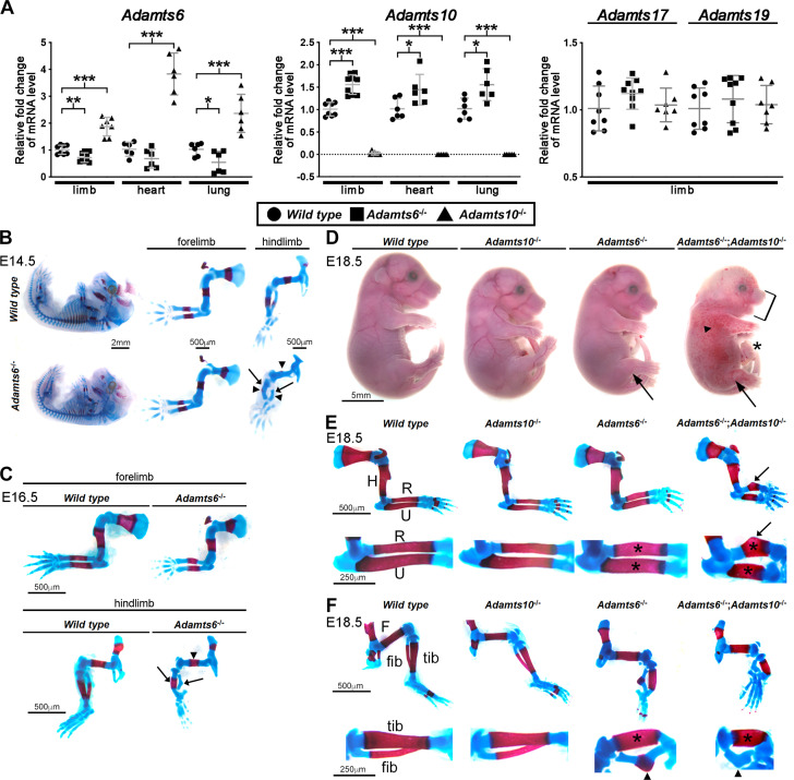
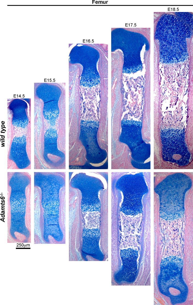
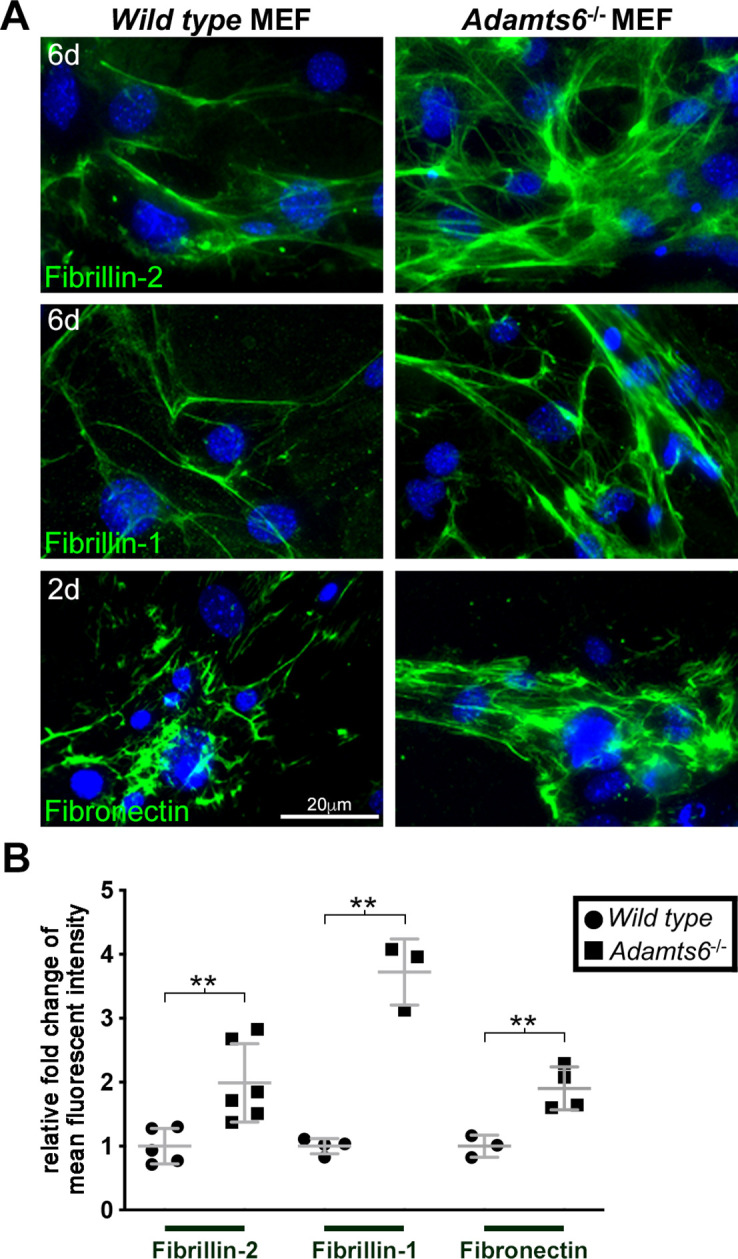
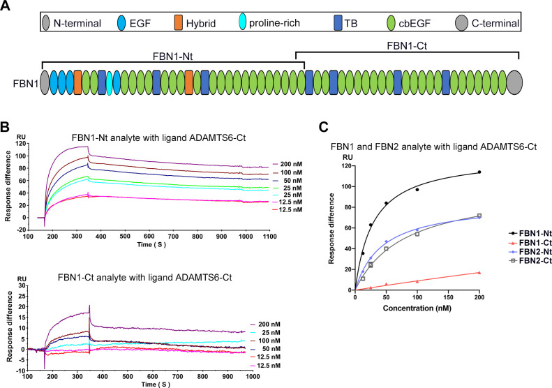
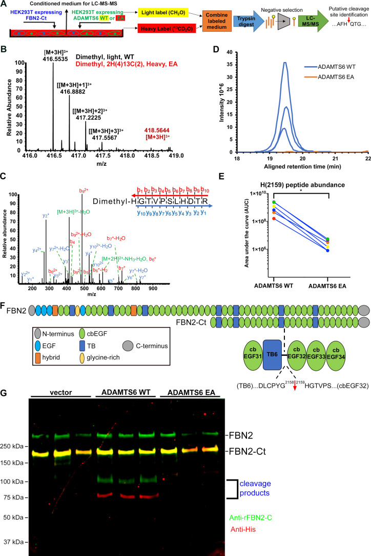
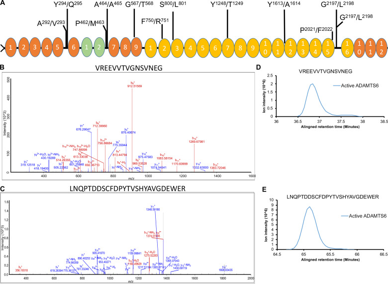
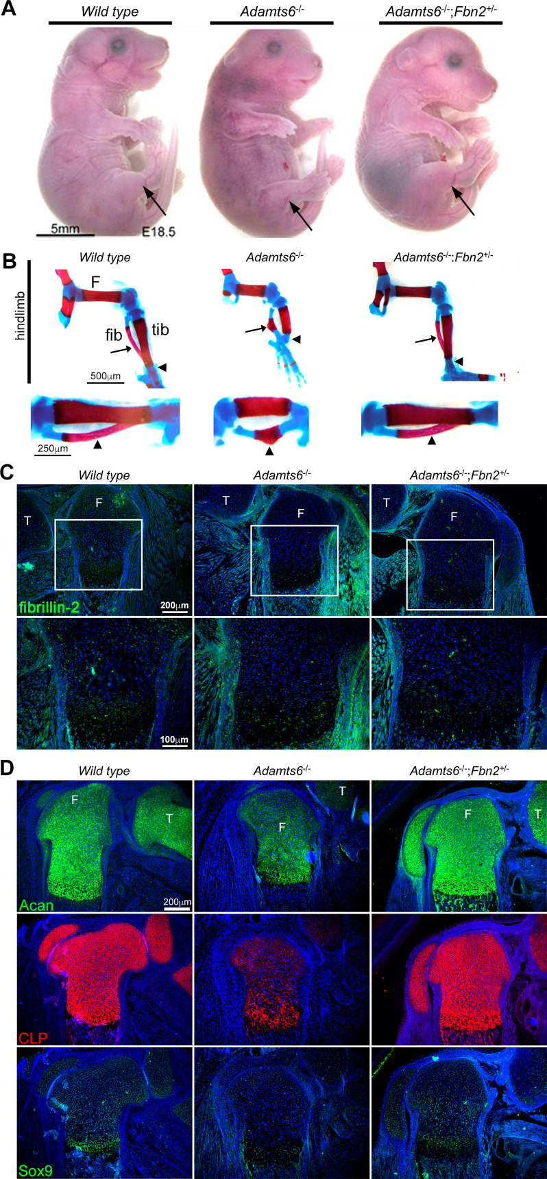
The embryonic extracellular matrix (ECM) undergoes transition to mature ECM as development progresses, yet few mechanisms ensuring ECM proteostasis during this period are known. Fibrillin microfibrils are macromolecular ECM complexes serving structural and regulatory roles. In mice, and encoding the major microfibrillar components, are strongly expressed during embryogenesis, but fibrillin-1 is the major component observed in adult tissue microfibrils. Here, analysis of and mutant mouse embryos, lacking these homologous secreted metalloproteases individually and in combination, along with in vitro analysis of microfibrils, measurement of ADAMTS6-fibrillin affinities and N-terminomics discovery of ADAMTS6-cleaved sites, identifies a proteostatic mechanism contributing to postnatal fibrillin-2 reduction and fibrillin-1 dominance. The lack of ADAMTS6, alone and in combination with ADAMTS10 led to excess fibrillin-2 in perichondrium, with impaired skeletal development defined by a drastic reduction of aggrecan and cartilage link protein, impaired BMP signaling in cartilage, and increased GDF5 sequestration in fibrillin-2-rich tissue. Although ADAMTS6 cleaves fibrillin-1 and fibrillin-2 as well as fibronectin, which provides the initial scaffold for microfibril assembly, primacy of the protease-substrate relationship between ADAMTS6 and fibrillin-2 was unequivocally established by reversal of the defects in embryos by genetic reduction of , but not .

摘要

胚胎细胞外基质 (ECM) 在发育过程中经历向成熟 ECM 的转变,但在此期间确保 ECM 蛋白稳态的机制知之甚少。纤维连接蛋白微纤维是具有结构和调节作用的大型 ECM 复合物。在小鼠中, 和 编码主要的微纤维成分,在胚胎发生过程中强烈表达,但在成年组织微纤维中观察到的主要成分是纤维连接蛋白 1。在这里,分析 和 突变体小鼠胚胎,单独和组合缺失这些同源分泌金属蛋白酶,以及体外微纤维分析、ADAMTS6-纤维连接蛋白亲和力的测量和 ADAMTS6 切割位点的 N 端组学发现,确定了一种有助于出生后纤维连接蛋白 2 减少和纤维连接蛋白 1 优势的蛋白稳态机制。单独缺乏 ADAMTS6 以及与 ADAMTS10 组合缺乏导致软骨膜中纤维连接蛋白 2 过多,软骨发育受损定义为聚集蛋白聚糖和软骨连接蛋白严重减少、软骨中 BMP 信号受损以及富含纤维连接蛋白 2 的组织中 GDF5 被隔离。尽管 ADAMTS6 切割纤维连接蛋白 1 和纤维连接蛋白 2 以及纤维连接蛋白,纤维连接蛋白为微纤维组装提供初始支架,但通过遗传减少 逆转 胚胎中的缺陷,明确确立了 ADAMTS6 和纤维连接蛋白 2 之间蛋白酶-底物关系的优先级,而不是 。

https://cdn.ncbi.nlm.nih.gov/pmc/blobs/f3ce/9064305/77bde6f4d2ff/elife-71142-fig11.jpg
https://cdn.ncbi.nlm.nih.gov/pmc/blobs/f3ce/9064305/550cfce463b4/elife-71142-fig1.jpg
https://cdn.ncbi.nlm.nih.gov/pmc/blobs/f3ce/9064305/9114031f76fd/elife-71142-fig1-figsupp1.jpg
https://cdn.ncbi.nlm.nih.gov/pmc/blobs/f3ce/9064305/eb502c91fe89/elife-71142-fig1-figsupp2.jpg
https://cdn.ncbi.nlm.nih.gov/pmc/blobs/f3ce/9064305/83e772360c84/elife-71142-fig1-figsupp3.jpg
https://cdn.ncbi.nlm.nih.gov/pmc/blobs/f3ce/9064305/128dca3eb478/elife-71142-fig1-figsupp4.jpg
https://cdn.ncbi.nlm.nih.gov/pmc/blobs/f3ce/9064305/75f62bd9a520/elife-71142-fig1-figsupp5.jpg
https://cdn.ncbi.nlm.nih.gov/pmc/blobs/f3ce/9064305/827e6b336e16/elife-71142-fig1-figsupp6.jpg
https://cdn.ncbi.nlm.nih.gov/pmc/blobs/f3ce/9064305/25c42f9e1921/elife-71142-fig1-figsupp7.jpg
https://cdn.ncbi.nlm.nih.gov/pmc/blobs/f3ce/9064305/f0a48d50a3a9/elife-71142-fig1-figsupp8.jpg
https://cdn.ncbi.nlm.nih.gov/pmc/blobs/f3ce/9064305/34cce46286fc/elife-71142-fig1-figsupp9.jpg
https://cdn.ncbi.nlm.nih.gov/pmc/blobs/f3ce/9064305/5ec284c8e35f/elife-71142-fig1-figsupp10.jpg
https://cdn.ncbi.nlm.nih.gov/pmc/blobs/f3ce/9064305/a82b5f4d4612/elife-71142-fig2.jpg
https://cdn.ncbi.nlm.nih.gov/pmc/blobs/f3ce/9064305/2818a6d4743f/elife-71142-fig3.jpg
https://cdn.ncbi.nlm.nih.gov/pmc/blobs/f3ce/9064305/f8a89055c459/elife-71142-fig3-figsupp1.jpg
https://cdn.ncbi.nlm.nih.gov/pmc/blobs/f3ce/9064305/f16bdbf69f92/elife-71142-fig3-figsupp2.jpg
https://cdn.ncbi.nlm.nih.gov/pmc/blobs/f3ce/9064305/076e08fd7400/elife-71142-fig3-figsupp3.jpg
https://cdn.ncbi.nlm.nih.gov/pmc/blobs/f3ce/9064305/9eaa70af6579/elife-71142-fig3-figsupp4.jpg
https://cdn.ncbi.nlm.nih.gov/pmc/blobs/f3ce/9064305/09ba79b3ffe3/elife-71142-fig4.jpg
https://cdn.ncbi.nlm.nih.gov/pmc/blobs/f3ce/9064305/bc5f32f7d0fc/elife-71142-fig4-figsupp1.jpg
https://cdn.ncbi.nlm.nih.gov/pmc/blobs/f3ce/9064305/a8ab59f8bba5/elife-71142-fig5.jpg
https://cdn.ncbi.nlm.nih.gov/pmc/blobs/f3ce/9064305/feb25c291097/elife-71142-fig5-figsupp1.jpg
https://cdn.ncbi.nlm.nih.gov/pmc/blobs/f3ce/9064305/e327deac995d/elife-71142-fig6.jpg
https://cdn.ncbi.nlm.nih.gov/pmc/blobs/f3ce/9064305/80da2b315f48/elife-71142-fig6-figsupp1.jpg
https://cdn.ncbi.nlm.nih.gov/pmc/blobs/f3ce/9064305/346b0212617f/elife-71142-fig7.jpg
https://cdn.ncbi.nlm.nih.gov/pmc/blobs/f3ce/9064305/1e0286b38e07/elife-71142-fig7-figsupp1.jpg
https://cdn.ncbi.nlm.nih.gov/pmc/blobs/f3ce/9064305/34b3042c604e/elife-71142-fig7-figsupp2.jpg
https://cdn.ncbi.nlm.nih.gov/pmc/blobs/f3ce/9064305/91326b56248f/elife-71142-fig8.jpg
https://cdn.ncbi.nlm.nih.gov/pmc/blobs/f3ce/9064305/c17363c3c296/elife-71142-fig8-figsupp1.jpg
https://cdn.ncbi.nlm.nih.gov/pmc/blobs/f3ce/9064305/c390c8d9f07c/elife-71142-fig8-figsupp2.jpg
https://cdn.ncbi.nlm.nih.gov/pmc/blobs/f3ce/9064305/02a344dca64e/elife-71142-fig8-figsupp3.jpg
https://cdn.ncbi.nlm.nih.gov/pmc/blobs/f3ce/9064305/9e4984fe64fc/elife-71142-fig8-figsupp4.jpg
https://cdn.ncbi.nlm.nih.gov/pmc/blobs/f3ce/9064305/ed6af388e52e/elife-71142-fig8-figsupp5.jpg
https://cdn.ncbi.nlm.nih.gov/pmc/blobs/f3ce/9064305/589a80dc6503/elife-71142-fig9.jpg
https://cdn.ncbi.nlm.nih.gov/pmc/blobs/f3ce/9064305/6e84ea859a45/elife-71142-fig10.jpg
https://cdn.ncbi.nlm.nih.gov/pmc/blobs/f3ce/9064305/d2216f68d620/elife-71142-fig10-figsupp1.jpg
https://cdn.ncbi.nlm.nih.gov/pmc/blobs/f3ce/9064305/77bde6f4d2ff/elife-71142-fig11.jpg

相似文献

1
Proteolysis of fibrillin-2 microfibrils is essential for normal skeletal development.原纤维蛋白-2 微纤维的蛋白水解对于正常骨骼发育是必不可少的。
Elife. 2022 May 3;11:e71142. doi: 10.7554/eLife.71142.
2
Adamts10 inactivation in mice leads to persistence of ocular microfibrils subsequent to reduced fibrillin-2 cleavage.在小鼠中敲除 Adamts10 会导致微纤维的持续存在,随后纤维连接蛋白-2 的裂解减少。
Matrix Biol. 2019 Apr;77:117-128. doi: 10.1016/j.matbio.2018.09.004. Epub 2018 Sep 7.
3
Unusual life cycle and impact on microfibril assembly of ADAMTS17, a secreted metalloprotease mutated in genetic eye disease.ADAMTS17,一种在遗传性眼病中发生突变的分泌型金属蛋白酶,其不寻常的生命周期及其对微纤维组装的影响。
Sci Rep. 2017 Feb 8;7:41871. doi: 10.1038/srep41871.
4
Limb- and tendon-specific Adamtsl2 deletion identifies a role for ADAMTSL2 in tendon growth in a mouse model for geleophysic dysplasia.肢体和肌腱特异性 Adamtsl2 缺失鉴定 ADAMTSL2 在 Geleophysic 发育不良小鼠模型中肌腱生长中的作用。
Matrix Biol. 2019 Sep;82:38-53. doi: 10.1016/j.matbio.2019.02.001. Epub 2019 Feb 7.
5
The ADAMTS/Fibrillin Connection: Insights into the Biological Functions of ADAMTS10 and ADAMTS17 and Their Respective Sister Proteases.ADAMTS/Fibrillin 连接:对 ADAMTS10 和 ADAMTS17 及其各自的姊妹蛋白酶的生物学功能的深入了解。
Biomolecules. 2020 Apr 12;10(4):596. doi: 10.3390/biom10040596.
6
Altered Ocular Fibrillin Microfibril Composition in Mice With a Glaucoma-Causing Mutation of Adamts10.导致青光眼的 Adamts10 基因突变小鼠的眼部纤维连接蛋白微纤维组成改变。
Invest Ophthalmol Vis Sci. 2021 Aug 2;62(10):26. doi: 10.1167/iovs.62.10.26.
7
Adamtsl2 deletion results in bronchial fibrillin microfibril accumulation and bronchial epithelial dysplasia--a novel mouse model providing insights into geleophysic dysplasia.Adamtsl2基因缺失导致支气管原纤维微原纤维积累和支气管上皮发育异常——一种为研究 geleophysic 发育异常提供见解的新型小鼠模型。
Dis Model Mech. 2015 May;8(5):487-99. doi: 10.1242/dmm.017046. Epub 2015 Mar 11.
8
ADAMTS6 cleaves the large latent TGFβ complex and increases the mechanotension of cells to activate TGFβ.ADAMTS6 裂解大型潜伏 TGFβ 复合物并增加细胞的力学张力以激活 TGFβ。
Matrix Biol. 2022 Dec;114:18-34. doi: 10.1016/j.matbio.2022.11.001. Epub 2022 Nov 8.
9
Cell-Based Interaction Analysis of ADAMTS Proteases and ADAMTS-Like Proteins with Fibrillin Microfibrils.基于细胞的ADAMTS蛋白酶和类ADAMTS蛋白与原纤维微原纤维的相互作用分析
Methods Mol Biol. 2020;2043:195-206. doi: 10.1007/978-1-4939-9698-8_16.
10
ADAMTS proteins as modulators of microfibril formation and function.ADAMTS蛋白作为微原纤维形成和功能的调节因子。
Matrix Biol. 2015 Sep;47:34-43. doi: 10.1016/j.matbio.2015.05.004. Epub 2015 May 7.

引用本文的文献

1
Extracellular matrix proteins refine microenvironments for pancreatic organogenesis from induced pluripotent stem cell differentiation.细胞外基质蛋白可优化诱导多能干细胞分化形成胰腺器官的微环境。
Theranostics. 2025 Jan 13;15(6):2229-2249. doi: 10.7150/thno.104883. eCollection 2025.
2
FBN2 promotes the proliferation, mineralization, and differentiation of osteoblasts to accelerate fracture healing.FBN2促进成骨细胞的增殖、矿化和分化,以加速骨折愈合。
Sci Rep. 2025 Feb 9;15(1):4843. doi: 10.1038/s41598-025-89215-6.
3
Proteomic insights into the extracellular matrix: a focus on proteoforms and their implications in health and disease.

本文引用的文献

1
A new mouse mutant with cleavage-resistant versican and isoform-specific versican mutants demonstrate that proteolysis at the Glu-Ala peptide bond in the V1 isoform is essential for interdigital web regression.一种具有抗切割多功能蛋白聚糖的新型小鼠突变体和多功能蛋白聚糖异构体特异性突变体表明,V1异构体中Glu-Ala肽键处的蛋白水解对于指间蹼的消退至关重要。
Matrix Biol Plus. 2021 May 14;10:100064. doi: 10.1016/j.mbplus.2021.100064. eCollection 2021 Jun.
2
A new MMP-mediated prodomain cleavage mechanism to activate bone morphogenetic proteins from the extracellular matrix.一种新的 MMP 介导的原结构域切割机制,用于从细胞外基质中激活骨形态发生蛋白。
FASEB J. 2021 Mar;35(3):e21353. doi: 10.1096/fj.202001264R.
3
蛋白质组学对外细胞基质的研究进展:重点关注蛋白质形式及其在健康和疾病中的意义。
Expert Rev Proteomics. 2024 Nov;21(11):463-481. doi: 10.1080/14789450.2024.2427136. Epub 2024 Nov 15.
4
Significance of Fibrillin-1, Filamin A, MMP2 and SOX9 in Mitral Valve Pathology.纤维连接蛋白 1、细丝蛋白 A、MMP2 和 SOX9 在二尖瓣病变中的意义。
Int J Mol Sci. 2024 Aug 29;25(17):9410. doi: 10.3390/ijms25179410.
5
The functional evolution of collembolan Ubx on the regulation of abdominal appendage formation.弹尾目昆虫Ubx在腹部附肢形成调控方面的功能演化。
Dev Genes Evol. 2024 Dec;234(2):135-151. doi: 10.1007/s00427-024-00718-0. Epub 2024 Jul 9.
6
Combined genetic-pharmacologic inactivation of tightly linked ADAMTS proteases in temporally specific windows uncovers distinct roles for versican proteolysis and glypican-6 in cardiac development.在特定时间窗口内联合遗传药理学失活紧密连锁的 ADAMTS 蛋白酶,揭示了 versican 蛋白水解和 glypican-6 在心脏发育中的不同作用。
Matrix Biol. 2024 Aug;131:1-16. doi: 10.1016/j.matbio.2024.05.003. Epub 2024 May 13.
7
Role of non-canonical post-translational modifications in gastrointestinal tumors.非经典翻译后修饰在胃肠道肿瘤中的作用。
Cancer Cell Int. 2023 Sep 30;23(1):225. doi: 10.1186/s12935-023-03062-x.
8
Editorial: ADAM, ADAMTS and astacin proteases: Challenges and breakthroughs in the -Omics era-Volume II.社论:ADAM、ADAMTS与虾红素蛋白酶:组学时代的挑战与突破——第二卷
Front Mol Biosci. 2023 Apr 7;10:1172288. doi: 10.3389/fmolb.2023.1172288. eCollection 2023.
9
Utilizing Variants Identified with Multiple Genome-Wide Association Study Methods Optimizes Genomic Selection for Growth Traits in Pigs.利用多种全基因组关联研究方法鉴定出的变异优化猪生长性状的基因组选择
Animals (Basel). 2023 Feb 17;13(4):722. doi: 10.3390/ani13040722.
10
Lack of evidence that fibrillin1 regulates bone morphogenetic protein 4 activity in kidney or lung.缺乏证据表明原纤维蛋白 1 可调节肾脏或肺部中的骨形态发生蛋白 4 活性。
Dev Dyn. 2023 Jun;252(6):761-769. doi: 10.1002/dvdy.578. Epub 2023 Mar 6.
A novel RGB-trichrome staining method for routine histological analysis of musculoskeletal tissues.
一种用于肌肉骨骼组织常规组织学分析的新型 RGB 三原色染色方法。
Sci Rep. 2020 Oct 7;10(1):16659. doi: 10.1038/s41598-020-74031-x.
4
Transcriptional adaptation: a mechanism underlying genetic robustness.转录适应:遗传稳健性的一种机制。
Development. 2020 Aug 14;147(15):dev186452. doi: 10.1242/dev.186452.
5
Proteomics identifies a convergent innate response to infective endocarditis and extensive proteolysis in vegetation components.蛋白质组学鉴定出感染性心内膜炎的固有反应和赘生物成分中广泛的蛋白水解作用存在趋同现象。
JCI Insight. 2020 Jul 23;5(14):135317. doi: 10.1172/jci.insight.135317.
6
The ADAMTS/Fibrillin Connection: Insights into the Biological Functions of ADAMTS10 and ADAMTS17 and Their Respective Sister Proteases.ADAMTS/Fibrillin 连接:对 ADAMTS10 和 ADAMTS17 及其各自的姊妹蛋白酶的生物学功能的深入了解。
Biomolecules. 2020 Apr 12;10(4):596. doi: 10.3390/biom10040596.
7
Elastic fiber ultrastructure and assembly.弹性纤维的超微结构与组装。
Matrix Biol. 2019 Nov;84:31-40. doi: 10.1016/j.matbio.2019.10.002. Epub 2019 Oct 24.
8
Alizarin Red and Alcian Blue Preparations to Visualize the Skeleton.茜素红和阿尔辛蓝制剂用于骨骼可视化。
Methods Mol Biol. 2020;2043:207-212. doi: 10.1007/978-1-4939-9698-8_17.
9
Expression Analysis by RNAscope™ In Situ Hybridization.使用RNAscope™原位杂交技术进行表达分析。
Methods Mol Biol. 2020;2043:173-178. doi: 10.1007/978-1-4939-9698-8_14.
10
Recent updates on the molecular network of elastic fiber formation.弹性纤维形成的分子网络的最新进展。
Essays Biochem. 2019 Sep 13;63(3):365-376. doi: 10.1042/EBC20180052.