Department of Neuroradiology, University Hospital Heidelberg, Heidelberg, Germany.
Department of Diagnostic and Interventional Radiology, University Hospital Heidelberg, Heidelberg, Germany.
J Neurointerv Surg. 2023 Jun;15(6):594-599. doi: 10.1136/neurintsurg-2022-018958. Epub 2022 May 4.
Endovascular embolization using liquid embolic agents (LEAs) is frequently applied for the treatment of intracranial vascular malformations. Appropriate visibility of LEAs during embolization is essential for visual control and to prevent complications. Since LEAs contain different radiopaque components of varying concentrations, our aim was the systematic assessment of the visibility of the most used LEAs in fluoroscopy.
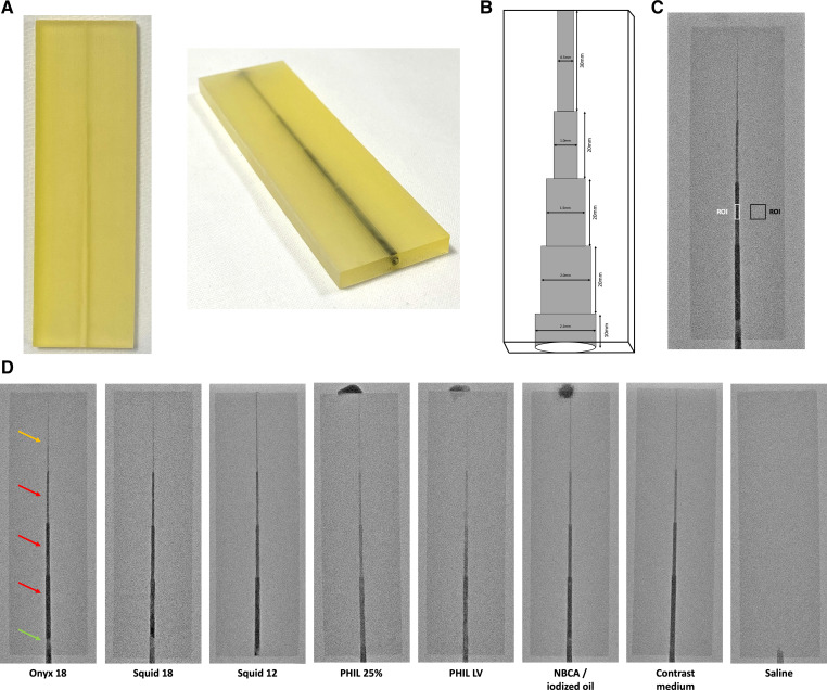
A specifically designed in vitro model, resembling cerebral vessels, was embolized with Onyx 18, Squid 18, Squid 12, PHIL (precipitating hydrophobic injectable liquid) 25%, PHIL LV (low viscosity) and NBCA (n-butyl cyanoacrylate) mixed with iodized oil (n=3 for each LEA), as well as with contrast medium and saline, both serving as a reference. Fluoroscopic image acquisition was performed in accordance with clinical routine settings. Visibility was graded quantitatively (contrast to noise ratio, CNR) and qualitatively (five-point scale).
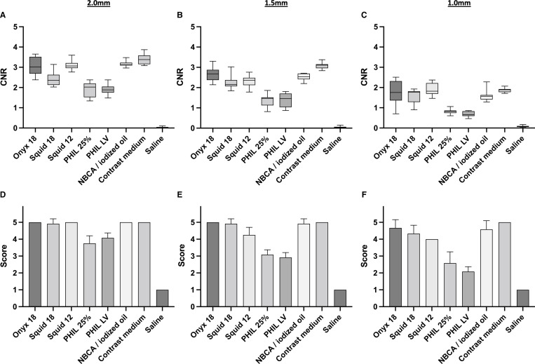
Overall, all LEAs provided at least acceptable visibility in this in vitro model. Onyx and Squid as well as NBCA mixed with iodized oil were best visible at a comparable level and superior to the formulations of PHIL, which did not differ in quantitative and qualitative analyses (eg, Onyx 18 vs PHIL 25% along the 2.0 mm sector: mean CNR±SD: 3.02±0.42 vs 1.92±0.35; mean score±SD: 5.00±0.00 vs 3.75±0.45; p≤0.001, respectively).
In this systematic in vitro study, relevant differences in the fluoroscopic visibility of LEAs in neurointerventional embolization procedures were demonstrated, while all investigated LEAs provided acceptable visibility in our in vitro model.
血管内栓塞治疗颅内血管畸形常采用液体栓塞剂(LEA)。栓塞过程中 LEA 的适当显影对于视觉控制和预防并发症至关重要。由于 LEA 含有不同浓度的不同不透射线成分,我们的目的是系统评估在透视术中最常用的 LEA 的可视性。
使用专门设计的类似于脑血管的体外模型,用 Onyx 18、Squid 18、Squid 12、PHIL(沉淀疏水性可注射液体)25%、PHIL LV(低粘度)和 NBCA(正丁基氰基丙烯酸酯)与碘油混合(每种 LEA 各 3 个样本),以及与对比剂和生理盐水一起进行栓塞,两者均作为参考。按照临床常规设置进行荧光透视图像采集。通过定量(对比噪声比,CNR)和定性(五分制)对显影进行评分。
总体而言,所有 LEA 在这个体外模型中均提供了至少可接受的可视性。Onyx 和 Squid 以及与碘油混合的 NBCA 在可比水平上具有最佳可视性,优于 PHIL 制剂,在定量和定性分析中没有差异(例如,在 2.0 毫米扇区中 Onyx 18 与 PHIL 25%:平均 CNR±SD:3.02±0.42 与 1.92±0.35;平均评分±SD:5.00±0.00 与 3.75±0.45;p≤0.001)。
在这项系统的体外研究中,展示了神经介入栓塞术中 LEA 荧光透视可视性的相关差异,而所有研究的 LEA 在我们的体外模型中均提供了可接受的可视性。