Laboratory of Experimental Pathology, Gonçalo Moniz Institute, Salvador, Bahia, Brazil.
Research Center on Mammary Oncology NPqOM/HOSPMEV, Federal University of Bahia, Salvador, Bahia, Brazil.
PLoS One. 2022 May 5;17(5):e0267648. doi: 10.1371/journal.pone.0267648. eCollection 2022.
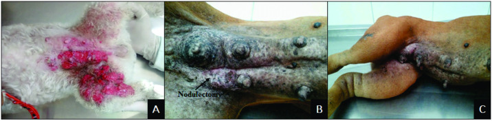
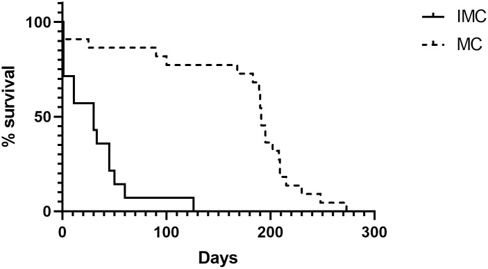
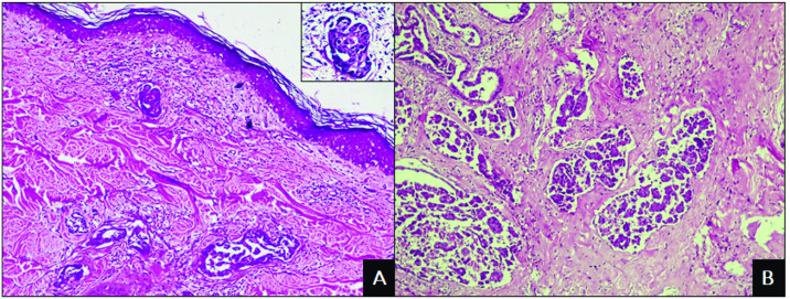
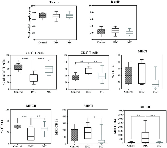
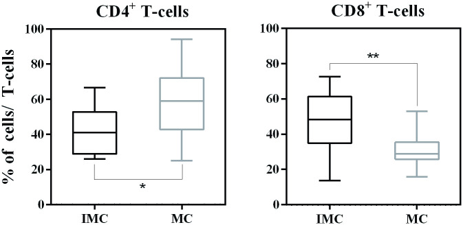
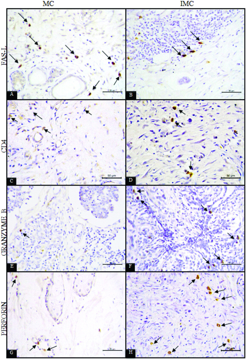
Inflammatory mammary carcinoma (IMC), a neoplasia affecting women and female dogs, is considered an aggressive cancer with high metastatic potential and a low survival rate. Studies focused on the tumour microenvironment indicate that the aggressive behaviour of this tumour is primarily correlated with immunological factors as well as inflammation. The objective of this study was to analyse the possible strategies used by the tumour cells to suppress the immune response in female dogs with IMC. Forty-six female dogs were divided into three groups: control (C, n = 10), IMC (n = 14) and mammary carcinoma (MC, n = 22). Clinical-pathological evaluations, survival at follow-up, immunophenotyping of leukocytes in peripheral blood and tumours, and immunohistochemical evaluation of CD4+, granzyme B, perforin and FAS-L were performed. Clinical and pathological results showed a higher frequency of the primary form of neoplasia, solid arrays of tumor cells and a lower survival rate in the IMC group (30 days). Morphometric analysis of inflammatory infiltrate revealed more lymphocytes and macrophages in the IMC group. Immunophenotyping analysis of peripheral blood revealed a higher frequency of CD8+ T-cells (p = 0.0017), a lower frequency of CD4+ T-cells (p <0.0001), and significantly higher mean MHCI and MHCII CD14+ fluorescence intensity in the IMC group (p = 0.038 and p = 0.0117, respectively). The immunohistochemical evaluation of tumour sections showed fewer FAS-L-positive inflammatory cells in the IMC group. These results suggest the important contribution of CD8+ T-cells, macrophages and FAS-L in the aggressiveness of IMC.
炎性乳腺癌(IMC)是一种影响女性和雌性犬的肿瘤,被认为是一种具有高转移潜能和低生存率的侵袭性癌症。针对肿瘤微环境的研究表明,这种肿瘤的侵袭性行为主要与免疫因素和炎症有关。本研究旨在分析 IMC 雌性犬肿瘤细胞抑制免疫反应的可能策略。46 只雌性犬分为三组:对照组(C,n=10)、IMC 组(n=14)和乳腺癌组(MC,n=22)。进行临床病理评估、随访生存情况、外周血和肿瘤白细胞免疫表型分析以及 CD4+、颗粒酶 B、穿孔素和 FAS-L 的免疫组化评价。临床和病理结果显示,IMC 组原发性肿瘤的发生频率更高,肿瘤细胞呈实性排列,生存率更低(30 天)。炎性浸润的形态计量分析显示 IMC 组淋巴细胞和巨噬细胞较多。外周血免疫表型分析显示,CD8+T 细胞频率更高(p=0.0017),CD4+T 细胞频率更低(p<0.0001),IMC 组平均 MHCI 和 MHCII CD14+荧光强度显著更高(p=0.038 和 p=0.0117)。肿瘤切片的免疫组化评价显示,IMC 组 FAS-L 阳性炎性细胞较少。这些结果表明 CD8+T 细胞、巨噬细胞和 FAS-L 在 IMC 的侵袭性中具有重要作用。