• 文献检索
  • 文档翻译
  • 深度研究
  • 学术资讯
  • Suppr Zotero 插件Zotero 插件
  • 邀请有礼
  • 套餐&价格
  • 历史记录
应用&插件
Suppr Zotero 插件Zotero 插件浏览器插件Mac 客户端Windows 客户端微信小程序
定价
高级版会员购买积分包购买API积分包
服务
文献检索文档翻译深度研究API 文档MCP 服务
关于我们
关于 Suppr公司介绍联系我们用户协议隐私条款
关注我们

Suppr 超能文献

核心技术专利:CN118964589B侵权必究
粤ICP备2023148730 号-1Suppr @ 2026

文献检索

告别复杂PubMed语法,用中文像聊天一样搜索,搜遍4000万医学文献。AI智能推荐,让科研检索更轻松。

立即免费搜索

文件翻译

保留排版,准确专业,支持PDF/Word/PPT等文件格式,支持 12+语言互译。

免费翻译文档

深度研究

AI帮你快速写综述,25分钟生成高质量综述,智能提取关键信息,辅助科研写作。

立即免费体验

COVID-19 幸存者在 6 个月随访时的定量吸气呼气胸部 CT 表现。

Quantitative inspiratory-expiratory chest CT findings in COVID-19 survivors at the 6-month follow-up.

机构信息

Department of Radiology, Union Hospital, Tongji Medical College, Huazhong University of Science and Technology, 1277 Jiefang Avenue, Wuhan, 43002, Hubei, People's Republic of China.

Hubei Province Key Laboratory of Molecular Imaging, Wuhan, 430022, People's Republic of China.

出版信息

Sci Rep. 2022 May 5;12(1):7402. doi: 10.1038/s41598-022-11237-1.

DOI:10.1038/s41598-022-11237-1
PMID:35513692
原文链接:https://pmc.ncbi.nlm.nih.gov/articles/PMC9070972/
Abstract

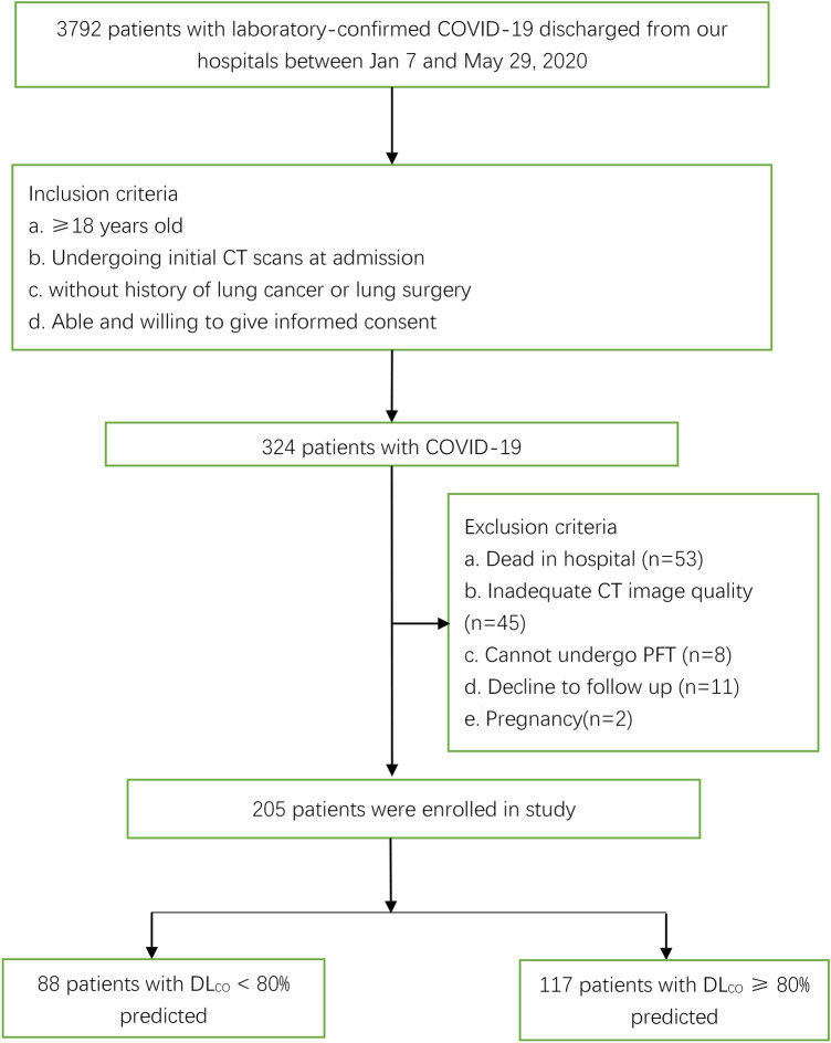
We evaluated pulmonary sequelae in COVID-19 survivors by quantitative inspiratory-expiratory chest CT (QCT) and explored abnormal pulmonary diffusion risk factors at the 6-month follow-up. This retrospective study enrolled 205 COVID-19 survivors with baseline CT data and QCT scans at 6-month follow-up. Patients without follow-up pulmonary function tests were excluded. All subjects were divided into group 1 (carbon monoxide diffusion capacity [DL] < 80% predicted, n = 88) and group 2 (DL ≥ 80% predicted, n = 117). Clinical characteristics and lung radiological changes were recorded. Semiquantitative total CT score (0-25) was calculated by adding five lobes scores (0-5) according to the range of lesion involvement (0: no involvement; 1: < 5%; 2: 5-25%; 3: 26-50%; 4: 51-75%; 5: > 75%). Data was analyzed by two-sample t-test, Spearman test, etc. 29% survivors showed air trapping by follow-up QCT. Semiquantitative CT score and QCT parameter of air trapping in group 1 were significantly greater than group 2 (p < 0.001). Decreased DL was negatively correlated with the follow-up CT score for ground-glass opacity (r = - 0.246, p = 0.003), reticulation (r = - 0.206, p = 0.002), air trapping (r = - 0.220, p = 0.002) and relative lung volume changes (r = - 0.265, p = 0.001). COVID-19 survivors with lung diffusion deficits at 6-month follow-up tended to develop air trapping, possibly due to small-airway impairment.

摘要

我们通过定量吸气呼气胸部 CT(QCT)评估 COVID-19 幸存者的肺部后遗症,并探讨了 6 个月随访时异常肺部弥散的危险因素。这项回顾性研究纳入了 205 名有基线 CT 数据和 6 个月随访 QCT 扫描的 COVID-19 幸存者。排除了没有随访肺功能测试的患者。所有患者均分为 1 组(一氧化碳弥散量[DL]<80%预计值,n=88)和 2 组(DL≥80%预计值,n=117)。记录临床特征和肺部影像学变化。通过将五个肺叶的分数(0-5)相加,计算半定量总 CT 评分(0-25),根据病变累及范围(0:无累及;1:<5%;2:5-25%;3:26-50%;4:51-75%;5:>75%)。采用两样本 t 检验、Spearman 检验等方法进行数据分析。29%的幸存者在随访 QCT 中显示空气潴留。1 组的半定量 CT 评分和 QCT 空气潴留参数明显大于 2 组(p<0.001)。DL 降低与随访 CT 评分的磨玻璃影(r=-0.246,p=0.003)、网状影(r=-0.206,p=0.002)、空气潴留(r=-0.220,p=0.002)和相对肺容量变化(r=-0.265,p=0.001)呈负相关。6 个月随访时肺弥散功能下降的 COVID-19 幸存者容易发生空气潴留,可能与小气道损伤有关。

https://cdn.ncbi.nlm.nih.gov/pmc/blobs/9037/9072427/f3787fa3aaf9/41598_2022_11237_Fig4_HTML.jpg
https://cdn.ncbi.nlm.nih.gov/pmc/blobs/9037/9072427/56d35ca61cf9/41598_2022_11237_Fig1_HTML.jpg
https://cdn.ncbi.nlm.nih.gov/pmc/blobs/9037/9072427/ce5f3de17291/41598_2022_11237_Fig2_HTML.jpg
https://cdn.ncbi.nlm.nih.gov/pmc/blobs/9037/9072427/60509ea7a47a/41598_2022_11237_Fig3_HTML.jpg
https://cdn.ncbi.nlm.nih.gov/pmc/blobs/9037/9072427/f3787fa3aaf9/41598_2022_11237_Fig4_HTML.jpg

相似文献

1
Quantitative inspiratory-expiratory chest CT findings in COVID-19 survivors at the 6-month follow-up.COVID-19 幸存者在 6 个月随访时的定量吸气呼气胸部 CT 表现。
Sci Rep. 2022 May 5;12(1):7402. doi: 10.1038/s41598-022-11237-1.
2
[Quantitative analysis of emphysema and air trapping at inspiratory and expiratory phase multi-slice spiral CT scan in smokers: correlation with pulmonary function test].吸烟者吸气和呼气期多层螺旋CT扫描对肺气肿和气陷的定量分析:与肺功能测试的相关性
Zhonghua Yi Xue Za Zhi. 2018 May 22;98(19):1467-1473. doi: 10.3760/cma.j.issn.0376-2491.2018.19.003.
3
Association of abnormal pulmonary vasculature on CT scan for COVID-19 infection with decreased diffusion capacity in follow up: A retrospective cohort study.COVID-19 感染 CT 扫描中异常肺血管与随访中弥散能力下降的相关性:一项回顾性队列研究。
PLoS One. 2021 Oct 15;16(10):e0257892. doi: 10.1371/journal.pone.0257892. eCollection 2021.
4
Air trapping in COVID-19 patients following hospital discharge: retrospective evaluation with paired inspiratory/expiratory thin-section CT.COVID-19 患者出院后患气腔滞留:采用配对吸气/呼气薄层 CT 的回顾性评估。
Eur Radiol. 2022 Jul;32(7):4427-4436. doi: 10.1007/s00330-022-08580-2. Epub 2022 Feb 28.
5
Post-discharge chest CT findings and pulmonary function tests in severe COVID-19 patients.重症新型冠状病毒肺炎患者出院后的胸部CT表现及肺功能测试
Eur J Radiol. 2021 May;138:109676. doi: 10.1016/j.ejrad.2021.109676. Epub 2021 Mar 20.
6
A Follow-Up Study of Lung Function and Chest Computed Tomography at 6 Months after Discharge in Patients with Coronavirus Disease 2019.新冠肺炎患者出院后 6 个月时的肺功能和胸部计算机断层扫描随访研究。
Can Respir J. 2021 Feb 13;2021:6692409. doi: 10.1155/2021/6692409. eCollection 2021.
7
Quantitative CT Analysis in Patients with Pulmonary Emphysema: Do Calculated Differences Between Full Inspiration and Expiration Correlate with Lung Function?定量 CT 分析在肺气肿患者中的应用:吸气末和呼气相的计算差值与肺功能相关吗?
Int J Chron Obstruct Pulmon Dis. 2020 Aug 3;15:1877-1886. doi: 10.2147/COPD.S253602. eCollection 2020.
8
Expiratory CT scans for chronic airway disease: correlation with pulmonary function test results.慢性气道疾病的呼气期CT扫描:与肺功能测试结果的相关性
AJR Am J Roentgenol. 1998 Feb;170(2):301-7. doi: 10.2214/ajr.170.2.9456933.
9
Post-Acute Sequelae of COVID-19 Pneumonia: Six-month Chest CT Follow-up.新型冠状病毒肺炎的急性后遗症:胸部CT六个月随访
Radiology. 2021 Nov;301(2):E396-E405. doi: 10.1148/radiol.2021210834. Epub 2021 Jul 27.
10
Evaluation of disease severity with quantitative chest CT in COVID-19 patients.评估 COVID-19 患者的疾病严重程度的定量胸部 CT。
Diagn Interv Radiol. 2021 Mar;27(2):164-171. doi: 10.5152/dir.2020.20281.

引用本文的文献

1
The relationship between the timing of lung surgery and postoperative pulmonary complications in patients after SARS-CoV-2 infection: a prospective cohort study.新型冠状病毒2感染后患者肺手术时机与术后肺部并发症的关系:一项前瞻性队列研究。
Front Med (Lausanne). 2025 Jul 28;12:1640475. doi: 10.3389/fmed.2025.1640475. eCollection 2025.
2
Long-Term Cardiopulmonary Function After COVID-19-Associated Acute Respiratory Distress Syndrome: A Multicenter Case-Control Study.新冠病毒相关急性呼吸窘迫综合征后的长期心肺功能:一项多中心病例对照研究
Crit Care Explor. 2025 Jul 10;7(7):e1286. doi: 10.1097/CCE.0000000000001286. eCollection 2025 Jul 1.
3

本文引用的文献

1
Post-Acute Sequelae of COVID-19 Pneumonia: Six-month Chest CT Follow-up.新型冠状病毒肺炎的急性后遗症:胸部CT六个月随访
Radiology. 2021 Nov;301(2):E396-E405. doi: 10.1148/radiol.2021210834. Epub 2021 Jul 27.
2
Radiological and functional lung sequelae of COVID-19: a systematic review and meta-analysis.COVID-19 的放射学和功能肺部后遗症:系统评价和荟萃分析。
BMC Pulm Med. 2021 Mar 22;21(1):97. doi: 10.1186/s12890-021-01463-0.
3
Quantitative CT imaging and advanced visualization methods: potential application in novel coronavirus disease 2019 (COVID-19) pneumonia.
FOT Technique Applied for Monitoring of COVID-19 Pneumonia Reveals Small Airways Involvement.
应用FOT技术监测新冠肺炎肺炎显示小气道受累。
Diagnostics (Basel). 2024 May 31;14(11):1160. doi: 10.3390/diagnostics14111160.
4
Pulmonary Function, Computed Tomography Lung Abnormalities, and Small Airway Disease after COVID-19: 3-, 6-, and 9-Month Follow-Up.新型冠状病毒肺炎后3、6和9个月随访时的肺功能、计算机断层扫描肺部异常及小气道疾病
J Clin Med. 2024 May 7;13(10):2733. doi: 10.3390/jcm13102733.
5
Chronic Lung Injury after COVID-19 Pneumonia: Clinical, Radiologic, and Histopathologic Perspectives.新冠肺炎后慢性肺损伤:临床、放射学和组织病理学视角。
Radiology. 2024 Jan;310(1):e231643. doi: 10.1148/radiol.231643.
6
Unraveling pathophysiologic mechanisms contributing to symptoms in patients with post-acute sequelae of COVID-19 (PASC): A retrospective study.解析导致 COVID-19 后急性后遗症(PASC)患者症状的病理生理机制:一项回顾性研究。
Physiol Rep. 2023 Jun;11(12):e15754. doi: 10.14814/phy2.15754.
7
Six-month follow-up after recovery of COVID-19 Delta variant survivors CT-based deep learning.新冠病毒德尔塔变异株康复者六个月随访:基于CT的深度学习
Front Med (Lausanne). 2023 Feb 2;10:1103559. doi: 10.3389/fmed.2023.1103559. eCollection 2023.
8
Longitudinal Assessment of Chest CT Findings and Pulmonary Function after COVID-19 Infection.COVID-19 感染后胸部 CT 表现和肺功能的纵向评估。
Radiology. 2023 Apr;307(2):e222888. doi: 10.1148/radiol.222888. Epub 2023 Feb 14.
9
Chronic Pulmonary Manifestations of COVID-19 Infection: Imaging Evaluation.COVID-19 感染的慢性肺部表现:影像学评估。
Radiology. 2023 Apr;307(2):e222379. doi: 10.1148/radiol.222379. Epub 2023 Jan 24.
10
Contrastive learning and subtyping of post-COVID-19 lung computed tomography images.新冠病毒感染后肺部计算机断层扫描图像的对比学习与亚型分析
Front Physiol. 2022 Oct 11;13:999263. doi: 10.3389/fphys.2022.999263. eCollection 2022.
定量CT成像与高级可视化方法:在2019年新型冠状病毒病(COVID-19)肺炎中的潜在应用
BJR Open. 2021 Jan 22;3(1):20200043. doi: 10.1259/bjro.20200043. eCollection 2021.
4
Short-term outpatient follow-up of COVID-19 patients: A multidisciplinary approach.COVID-19患者的短期门诊随访:多学科方法
EClinicalMedicine. 2021 Feb;32:100731. doi: 10.1016/j.eclinm.2021.100731. Epub 2021 Jan 28.
5
Six-month Follow-up Chest CT Findings after Severe COVID-19 Pneumonia.重症 COVID-19 肺炎患者 6 个月随访的胸部 CT 结果。
Radiology. 2021 Apr;299(1):E177-E186. doi: 10.1148/radiol.2021203153. Epub 2021 Jan 26.
6
Temporal changes of haematological and radiological findings of the COVID-19 infection-a review of literature.COVID-19 感染的血液学和影像学表现的时间变化——文献综述。
BMC Pulm Med. 2021 Jan 22;21(1):37. doi: 10.1186/s12890-020-01389-z.
7
Inspiratory and Expiratory Chest High-resolution CT: Small-airway Disease Evaluation in Patients with COVID-19.吸气和呼气胸部高分辨率 CT:COVID-19 患者的小气道疾病评估。
Curr Med Imaging. 2021;17(11):1299-1307. doi: 10.2174/1573405617999210112194621.
8
6-month consequences of COVID-19 in patients discharged from hospital: a cohort study.新冠肺炎出院患者 6 个月的后果:一项队列研究。
Lancet. 2021 Jan 16;397(10270):220-232. doi: 10.1016/S0140-6736(20)32656-8. Epub 2021 Jan 8.
9
Influenza virus infection increases ACE2 expression and shedding in human small airway epithelial cells.流感病毒感染增加人小气道上皮细胞中 ACE2 的表达和脱落。
Eur Respir J. 2021 Jul 1;58(1). doi: 10.1183/13993003.03988-2020. Print 2021 Jul.
10
Ultra-low-dose chest CT in adult patients with cystic fibrosis using a third-generation dual-source CT scanner.使用第三代双源CT扫描仪对成年囊性纤维化患者进行超低剂量胸部CT检查。
Radiol Med. 2021 Apr;126(4):544-552. doi: 10.1007/s11547-020-01304-w. Epub 2020 Nov 16.