Offroy Marc, Razafitianamaharavo Angelina, Beaussart Audrey, Pagnout Christophe, Duval Jérôme F L
Université de Lorraine, CNRS, LIEC F-54000 Nancy France
Université de Lorraine, CNRS, LIEC F-57000 Metz France.
RSC Adv. 2020 May 20;10(33):19258-19275. doi: 10.1039/d0ra00669f.
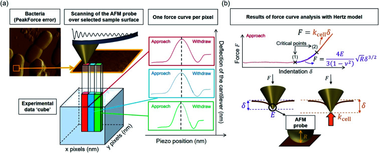
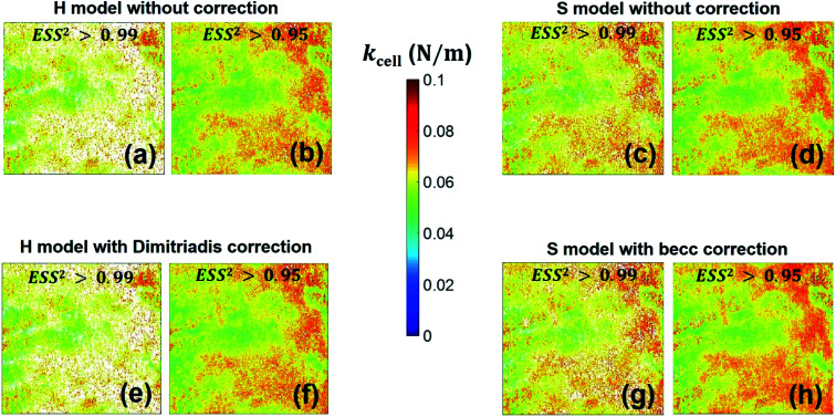
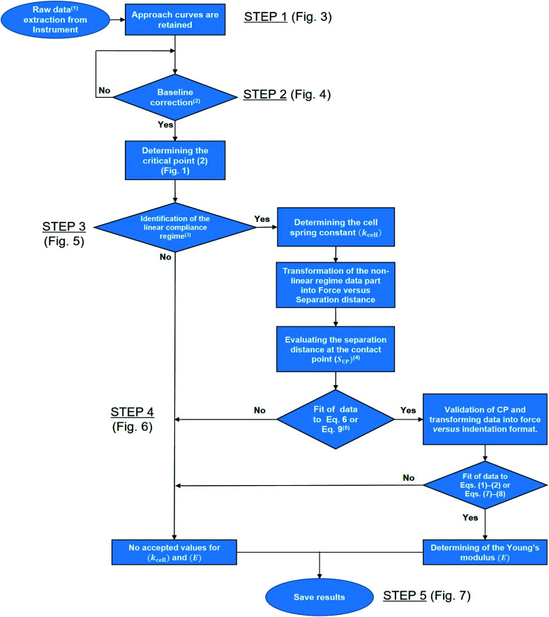
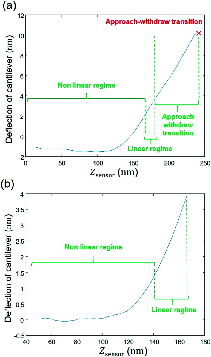
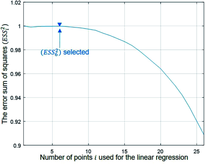
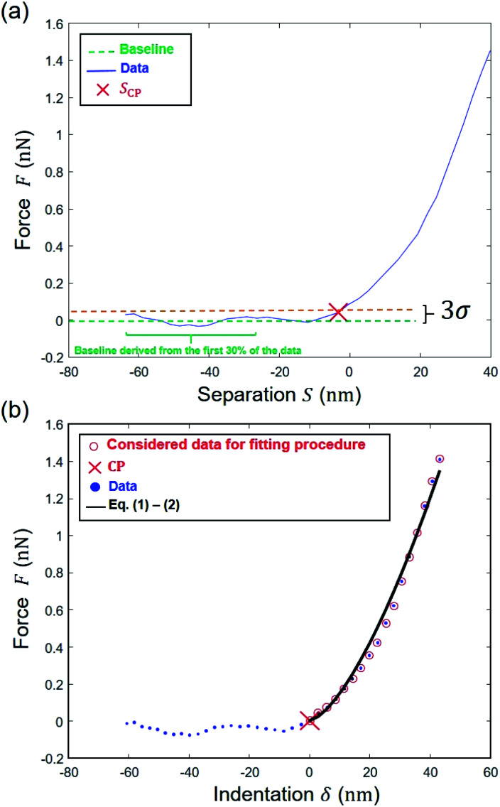
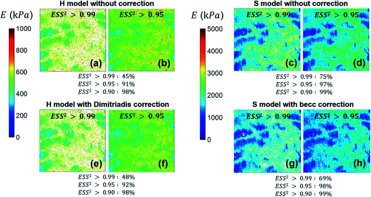
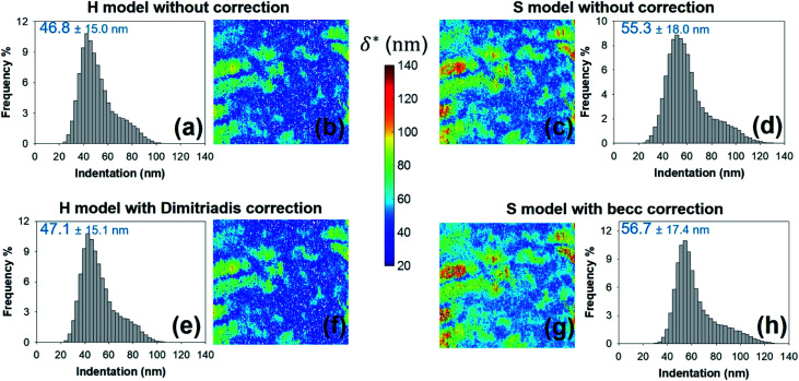
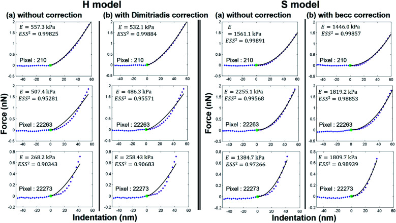
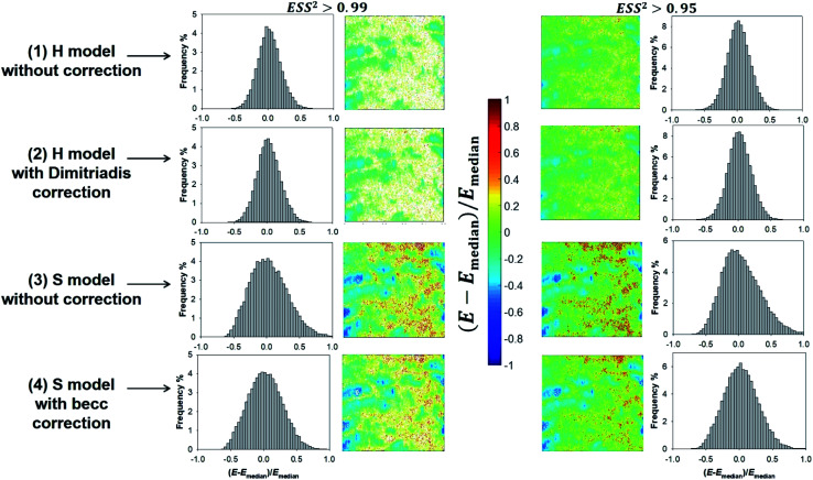
Atomic Force Microscopy (AFM) is a powerful technique for the measurement of mechanical properties of individual cells in two ( × ) or three ( × × time) dimensions. The instrumental progress makes it currently possible to generate a large amount of data in a relatively short time, which is particularly true for AFM operating in so-called PeakForce tapping mode (Bruker corporation). The latter corresponds to an AFM probe that periodically hits the sample surface while the pico-newton level interaction force is recorded from cantilever deflection. The method provides unprecedented high-resolution (a few tens of nm) imaging of the mechanical features of soft biological samples ( bacteria, yeasts) and of hard abiotic surfaces ( minerals). The rapid conversion of up to several tens of thousands spatially resolved force curves typically collected in AFM PeakForce tapping mode over a given cell surface area into comprehensive nanomechanical information requires the development of robust data analysis methodologies and dedicated numerical tools. In this work, we report an automated algorithm for (i) a rapid and unambiguous detection of the indentation regimes corresponding to non-linear and linear deformations of bacterial surfaces upon compression by the AFM probe, (ii) the subsequent evaluation of the Young modulus and cell surface stiffness, and (iii) the generation of spatial mappings of relevant nanomechanical properties at the single cell level. The procedure involves consistent evaluation of the contact point between the AFM probe and sample biosurface and that of the threshold indentation value marking the transition between non-linear and linear deformation regimes. For comparison purposes, the former regime is here analyzed on the basis of Hertz and Sneddon models corrected or not for effects of finite sample thickness. Analysis of AFM measurements performed on a selected strain is detailed to demonstrate the feasibility, rapidity and robustness of the here-proposed PeakForce data treatment process. The flexibility of the algorithm allows consideration of force curve parameterizations other than that detailed here, which may be desired for investigation of eukaryotes nanomechanics. The performance of the adopted Hertz-based and Sneddon-based contact mechanics formalisms in recovering experimental data and in identifying nanomechanical heterogeneities at the bacterium scale is further thoroughly discussed.
原子力显微镜(AFM)是一种强大的技术,可用于测量单个细胞在二维(×)或三维(××时间)维度上的力学性能。仪器的进步使得目前有可能在相对较短的时间内生成大量数据,对于在所谓的峰值力轻敲模式(布鲁克公司)下运行的AFM来说尤其如此。后者对应于一个AFM探针,它周期性地撞击样品表面,同时从悬臂梁的偏转记录皮牛级别的相互作用力。该方法为软生物样品(细菌、酵母)和硬非生物表面(矿物质)的力学特征提供了前所未有的高分辨率(几十纳米)成像。在AFM峰值力轻敲模式下,通常在给定的细胞表面积上收集多达数万条空间分辨的力曲线,并将其快速转换为全面的纳米力学信息,这需要开发强大的数据分析方法和专用的数值工具。在这项工作中,我们报告了一种自动化算法,用于(i)快速明确地检测与AFM探针压缩细菌表面时的非线性和线性变形相对应的压痕区域,(ii)随后评估杨氏模量和细胞表面刚度,以及(iii)在单细胞水平上生成相关纳米力学性能的空间映射。该过程涉及对AFM探针与样品生物表面之间的接触点以及标记非线性和线性变形区域之间过渡的阈值压痕值进行一致评估。为了进行比较,前者基于赫兹和斯内登模型进行分析,该模型针对有限样品厚度的影响进行了校正或未校正。详细分析了对选定菌株进行的AFM测量,以证明本文提出的峰值力数据处理过程的可行性、快速性和稳健性。该算法的灵活性允许考虑此处未详细说明的力曲线参数化,这可能是研究真核生物纳米力学所需要的。进一步深入讨论了所采用的基于赫兹和基于斯内登的接触力学形式在恢复实验数据和识别细菌尺度上的纳米力学异质性方面的性能。