Shimazaki Taishi, Deshpande Ameya, Hajra Anindya, Thomas Tijo, Muta Kyotaka, Yamada Naohito, Yasui Yuzo, Shoda Toshiyuki
Toxicology Research Laboratories, Central Pharmaceutical Research Institute, Japan Tobacco Inc., 1-13-2 Fukuura, Kanazawa-ku, Yokohama, Kanagawa 236-0004, Japan.
AIRA Matrix Private Limited, Dosti Pinnacle, 801, Rd Number 22, Wagle Industrial Estate, Thane, Maharashtra 400604, India.
J Toxicol Pathol. 2022 Apr;35(2):135-147. doi: 10.1293/tox.2021-0053. Epub 2021 Nov 27.
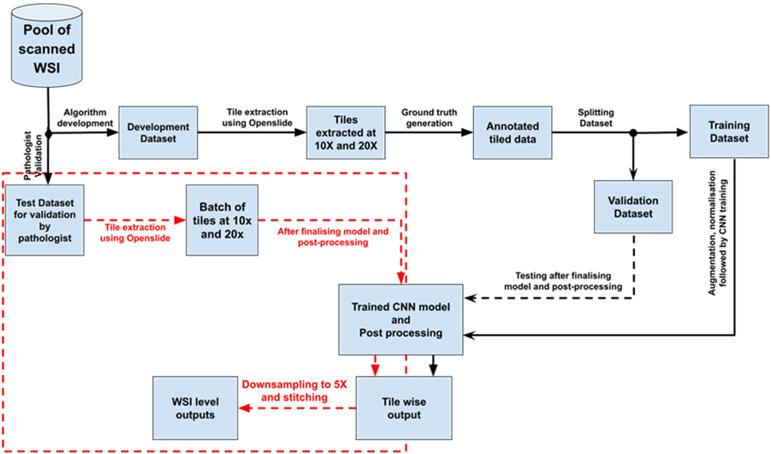
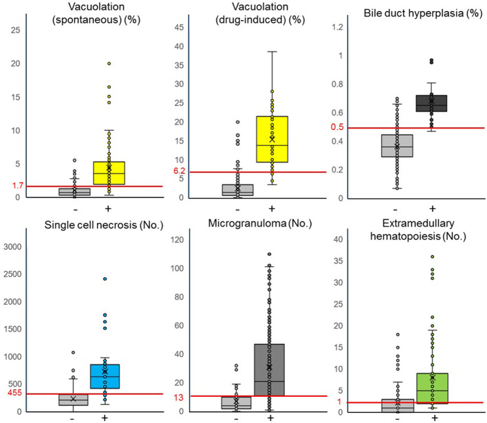
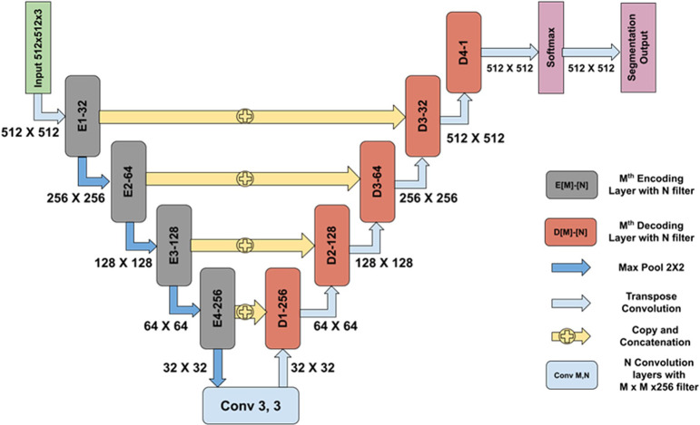
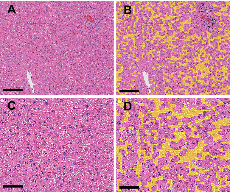
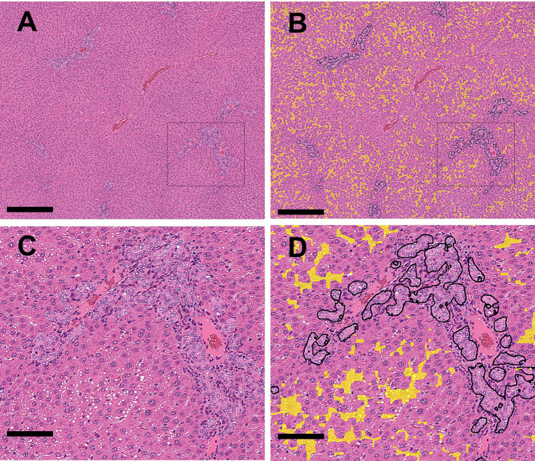
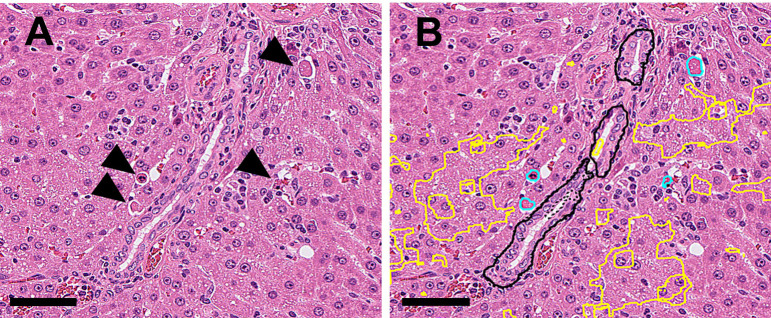
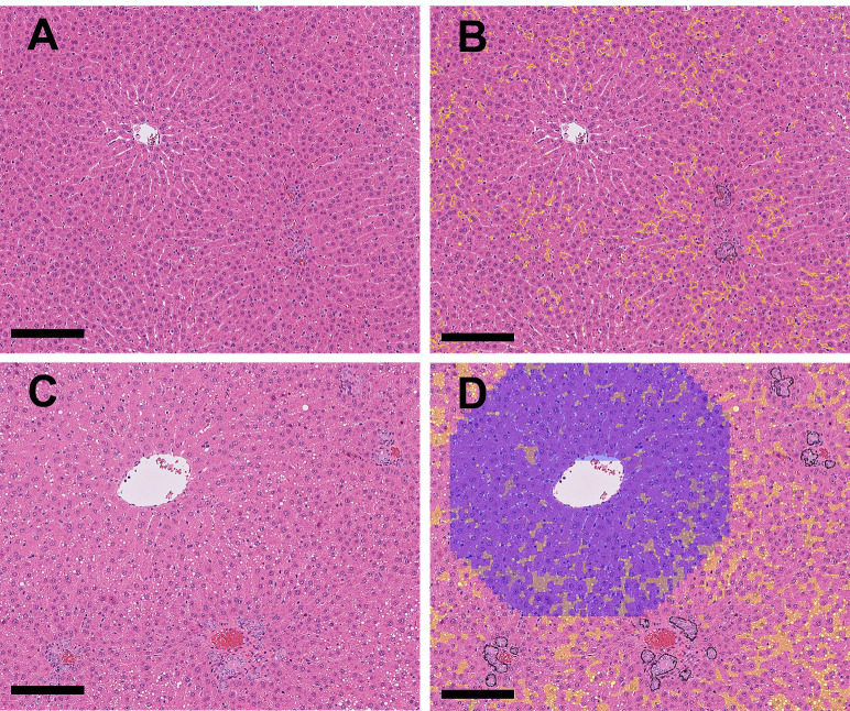
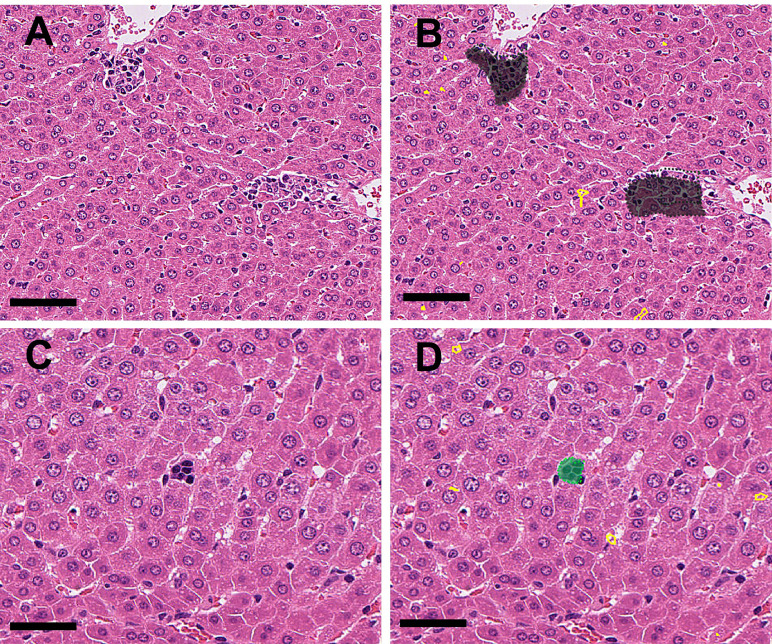
Artificial intelligence (AI)-based image analysis is increasingly being used for preclinical safety-assessment studies in the pharmaceutical industry. In this paper, we present an AI-based solution for preclinical toxicology studies. We trained a set of algorithms to learn and quantify multiple typical histopathological findings in whole slide images (WSIs) of the livers of young Sprague Dawley rats by using a U-Net-based deep learning network. The trained algorithms were validated using 255 liver WSIs to detect, classify, and quantify seven types of histopathological findings (including vacuolation, bile duct hyperplasia, and single-cell necrosis) in the liver. The algorithms showed consistently good performance in detecting abnormal areas. Approximately 75% of all specimens could be classified as true positive or true negative. In general, findings with clear boundaries with the surrounding normal structures, such as vacuolation and single-cell necrosis, were accurately detected with high statistical scores. The results of quantitative analyses and classification of the diagnosis based on the threshold values between "no findings" and "abnormal findings" correlated well with diagnoses made by professional pathologists. However, the scores for findings ambiguous boundaries, such as hepatocellular hypertrophy, were poor. These results suggest that deep learning-based algorithms can detect, classify, and quantify multiple findings simultaneously on rat liver WSIs. Thus, it can be a useful supportive tool for a histopathological evaluation, especially for primary screening in rat toxicity studies.
基于人工智能(AI)的图像分析在制药行业的临床前安全性评估研究中越来越多地被使用。在本文中,我们提出了一种用于临床前毒理学研究的基于AI的解决方案。我们训练了一组算法,通过使用基于U-Net的深度学习网络来学习和量化年轻Sprague Dawley大鼠肝脏全切片图像(WSIs)中的多种典型组织病理学发现。使用255张肝脏WSIs对训练好的算法进行验证,以检测、分类和量化肝脏中的七种组织病理学发现(包括空泡化、胆管增生和单细胞坏死)。这些算法在检测异常区域方面表现出始终如一的良好性能。大约75%的所有标本可以被分类为真阳性或真阴性。一般来说,与周围正常结构有清晰边界的发现,如空泡化和单细胞坏死,能够以高统计分数被准确检测到。基于“无发现”和“异常发现”之间阈值的定量分析和诊断分类结果与专业病理学家的诊断结果相关性良好。然而,对于边界模糊的发现,如肝细胞肥大,评分较差。这些结果表明,基于深度学习的算法可以在大鼠肝脏WSIs上同时检测、分类和量化多种发现。因此,它可以成为组织病理学评估的有用支持工具,特别是在大鼠毒性研究的初步筛选中。