Li Yang, Zhang Yu, Ding Jing-Li, Liu Ji-Chun, Xu Jian-Jun, Tang Yan-Hua, Yi Ying-Ping, Xu Wei-Chang, Yu Wen-Peng, Lu Chao, Yang Wei, Yang Jue-Sheng, Gong Yi, Zhou Jian-Liang
Department of Cardiovascular Surgery, The Second Affiliated Hospital of Nanchang University No. 1, Mingde Road Nanchang 330000 China
Department of Cardiovascular Surgery, Renji Hospital, School of Medicine, Shanghai Jiaotong University Shanghai China.
RSC Adv. 2019 Apr 16;9(21):11882-11893. doi: 10.1039/c9ra00408d. eCollection 2019 Apr 12.
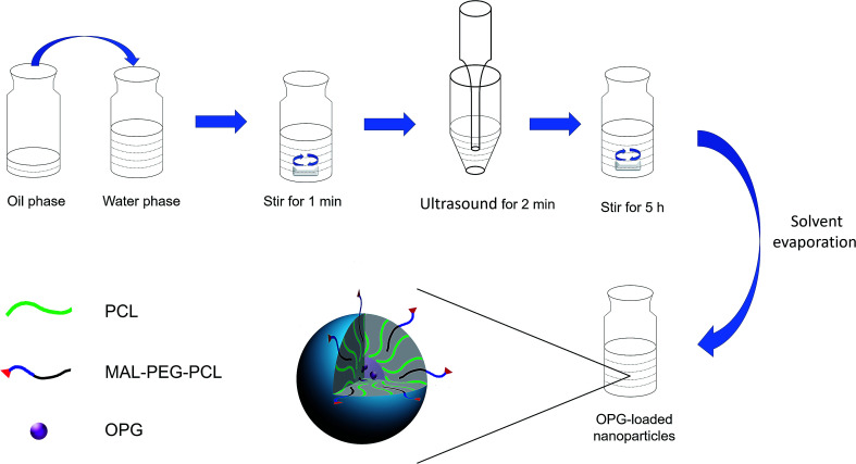
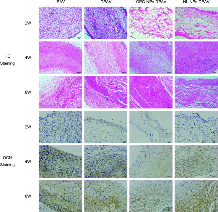
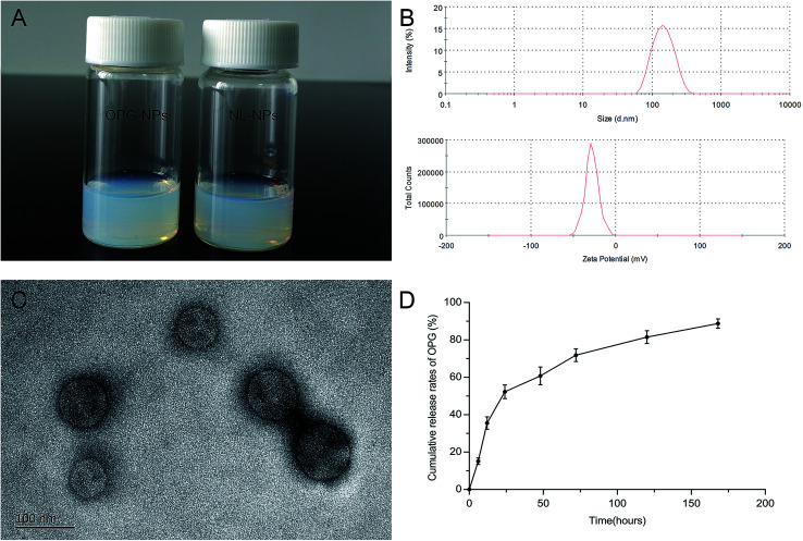
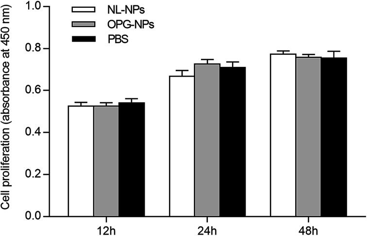
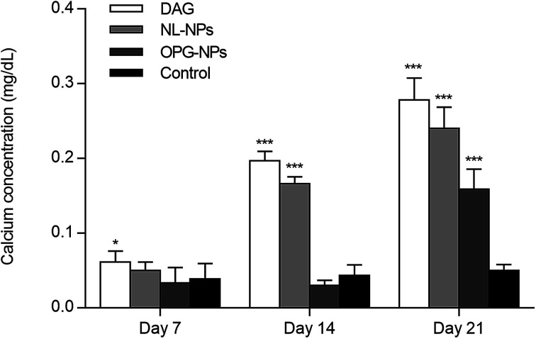
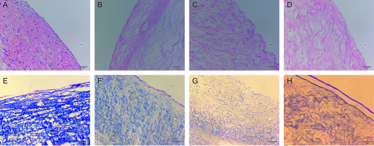
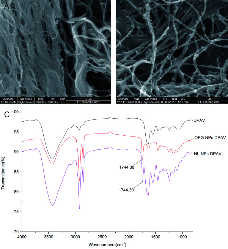
Decellularized valve stents are widely used in tissue-engineered heart valves because they maintain the morphological structure of natural valves, have good histocompatibility and low immunogenicity. However, the surface of the cell valve loses the original endothelial cell coverage, exposing collagen and causing calcification and decay of the valve in advance. In this study, poly ε-caprolactone (PCL) nanoparticles loaded with osteoprotegerin (OPG) were bridged to a decellularized valve using a nanoparticle drug delivery system and tissue engineering technology to construct a new anti-calcification composite valve with sustained release function. The PCL nanoparticles loaded with OPG were prepared an emulsion solvent evaporation method, which had a particle size of 133 nm and zeta potential of -27.8 mV. Transmission electron microscopy demonstrated that the prepared nanoparticles were round in shape, regular in size, and uniformly distributed, with an encapsulation efficiency of 75%, slow release , no burst release, no cytotoxicity to BMSCs, and contained OPG nanoparticles . There was a delay in the differentiation of BMSCs into osteoblasts. The decellularized valve modified by nanoparticles remained intact and its collagen fibers were continuous. After 8 weeks of subcutaneous implantation in rats, the morphological structure of the valve was almost complete, and the composite valve showed anti-calcification ability to a certain extent.
去细胞化瓣膜支架因其保留天然瓣膜的形态结构、具有良好的组织相容性和低免疫原性而被广泛应用于组织工程心脏瓣膜。然而,去细胞化瓣膜表面失去了原有的内皮细胞覆盖,暴露出胶原蛋白,导致瓣膜提前钙化和衰败。在本研究中,利用纳米颗粒药物递送系统和组织工程技术,将负载骨保护素(OPG)的聚ε-己内酯(PCL)纳米颗粒连接到去细胞化瓣膜上,构建一种具有缓释功能的新型抗钙化复合瓣膜。采用乳液溶剂蒸发法制备负载OPG的PCL纳米颗粒,其粒径为133nm,ζ电位为-27.8mV。透射电子显微镜显示,制备的纳米颗粒呈圆形,大小规则,分布均匀,包封率为75%,具有缓释效果,无突释现象,对骨髓间充质干细胞无细胞毒性,且含有OPG纳米颗粒。骨髓间充质干细胞向成骨细胞的分化存在延迟。纳米颗粒修饰的去细胞化瓣膜保持完整,其胶原纤维连续。大鼠皮下植入8周后,瓣膜的形态结构基本完整,复合瓣膜在一定程度上显示出抗钙化能力。