文献检索文档翻译深度研究
Suppr Zotero 插件Zotero 插件
邀请有礼套餐&价格历史记录

新学期,新优惠

限时优惠:9月1日-9月22日

30天高级会员仅需29元

1天体验卡首发特惠仅需5.99元

了解详情
不再提醒
插件&应用
Suppr Zotero 插件Zotero 插件浏览器插件Mac 客户端Windows 客户端微信小程序
高级版
套餐订阅购买积分包
AI 工具
文献检索文档翻译深度研究
关于我们
关于 Suppr公司介绍联系我们用户协议隐私条款
关注我们

Suppr 超能文献

核心技术专利:CN118964589B侵权必究
粤ICP备2023148730 号-1Suppr @ 2025

用作肿瘤靶向磁共振成像造影剂的肽修饰超小超顺磁性氧化铁的制备与表征

Preparation and characterization of peptide modified ultrasmall superparamagnetic iron oxides used as tumor targeting MRI contrast agent.

作者信息

Yin Jie, Yin Guangfu, Pu Ximing, Huang Zhongbing, Yao Dajin

机构信息

College of Materials Science and Engineering, Sichuan University No. 24, South 1st Section, 1st Ring Road Chengdu 610065 PR China

School of Automation and Information Engineering, Sichuan University of Science and Engineering Zigong 643000 PR China.

出版信息

RSC Adv. 2019 Jun 20;9(34):19397-19407. doi: 10.1039/c9ra02636c. eCollection 2019 Jun 19.


DOI:10.1039/c9ra02636c
PMID:35519366
原文链接:https://pmc.ncbi.nlm.nih.gov/articles/PMC9065400/
Abstract

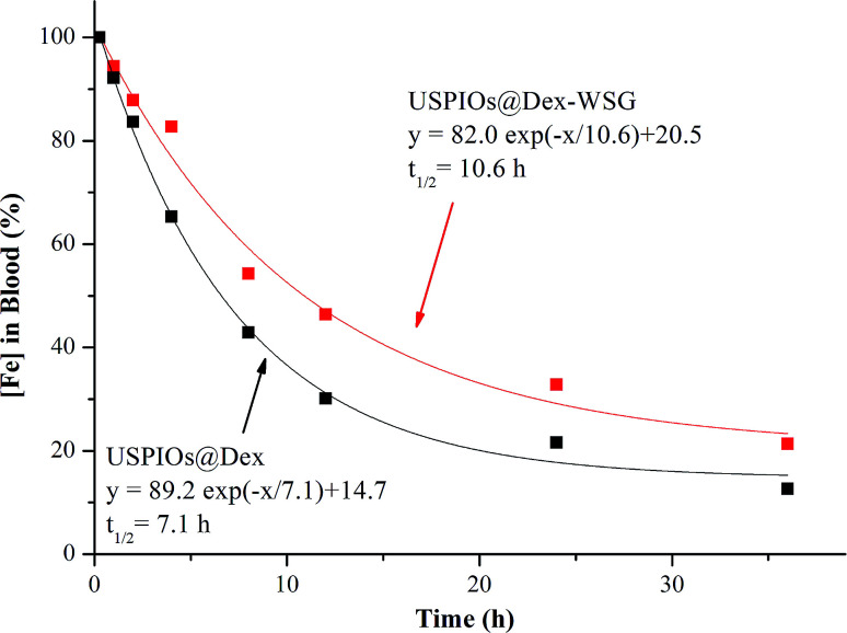
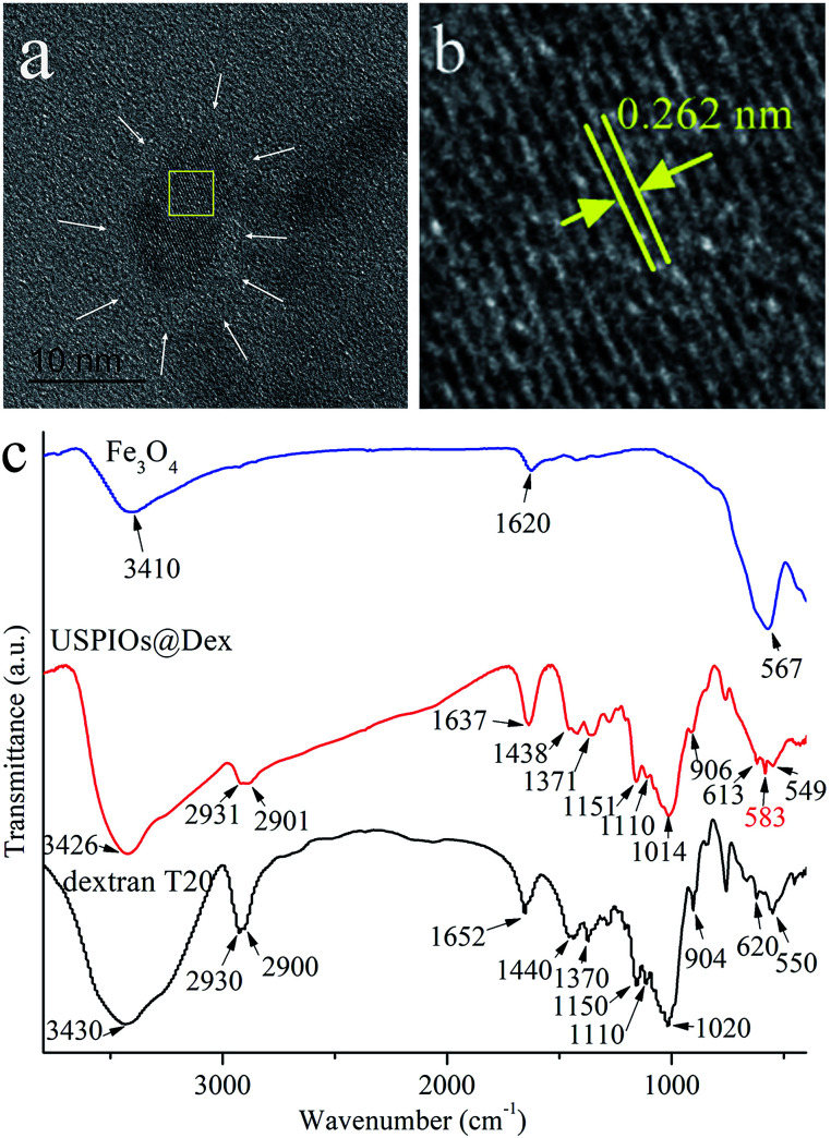
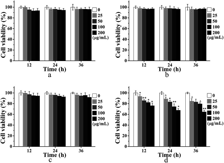
As desirable contrast agents for magnetic resonance imaging (MRI), ultrasmall superparamagnetic iron oxides (USPIOs) are required to exhibit both low cytotoxicity and specific targetability besides superparamagnetism to achieve better imaging contrast at lower dose, and cladding with biocompatible polymers and modification with targeting ligands are considered to be the most effective strategies. In this study, novel dextran wrapped and peptide WSGPGVWGASVK (peptide-WSG) grafted USPIOs were meticulously prepared and systematically characterized. Firstly, dextran (Dex) cladded USPIOs (USPIOs@Dex) were synthesized with a well-designed co-precipitation procedure in which the biocompatible dextran played dual roles of grain inhibitor and cladding agent. After that, sodium citrate was applied to carboxylize the hydroxyls of the dextran molecules an esterification reaction, and then tumor targeting peptide-WSG was grafted to the carboxyl groups by the EDC method. The XRD, TEM, and FTIR results showed that inverse spinel structure FeO crystallites were nucleated and grown in aqueous solution, and the catenulate dextran molecules gradually bound on their surface, meanwhile the growth of grains was inhibited. The size of original crystallite grains was about 7 nm, but the mean size of USPIOs@Dex aggregates was 165.20 nm. After surface modification by sodium citrate and peptide-WSG with ultrasonic agitation, the size of the USPIOs@Dex-WSG aggregates was smaller (66.06 nm) because the hydrophilicity was improved, so USPIOs@Dex-WSG could evade being eliminated by RES more easily, and prolong residence time in blood circulation. The VSM and T-weighted MRI results showed that USPIOs@Dex-WSG were superparamagnetic with a saturation magnetization of 44.65 emu g, and with high transverse relaxivity as the relaxivity coefficient value was 229.70 mM s. The results of MTT assays and the Prussian blue staining revealed that USPIOs@Dex-WSG exhibited nontoxicity for normal cells such as L929 and HUVECs, and were specifically targeted to the SKOV-3 cells. Thus, the novel dextran wrapped and WSG-peptide grafted USPIOs have potential to be applied as tumor active targeting contrast agents for MRI.

摘要

作为磁共振成像(MRI)理想的造影剂,除了超顺磁性外,超小超顺磁性氧化铁(USPIOs)还需要表现出低细胞毒性和特异性靶向性,以便在较低剂量下实现更好的成像对比度,而用生物相容性聚合物包覆和用靶向配体修饰被认为是最有效的策略。在本研究中,精心制备并系统表征了新型葡聚糖包裹且肽WSGPGVWGASVK(肽-WSG)接枝的USPIOs。首先,采用精心设计的共沉淀法合成了葡聚糖(Dex)包覆的USPIOs(USPIOs@Dex),其中生物相容性葡聚糖起到了晶粒抑制剂和包覆剂的双重作用。之后,通过酯化反应使用柠檬酸钠将葡聚糖分子的羟基羧基化,然后通过EDC法将肿瘤靶向肽-WSG接枝到羧基上。XRD、TEM和FTIR结果表明,反尖晶石结构的FeO微晶在水溶液中形核并生长,链状葡聚糖分子逐渐结合在其表面,同时晶粒生长受到抑制。原始微晶颗粒的尺寸约为7nm,但USPIOs@Dex聚集体的平均尺寸为165.20nm。经柠檬酸钠和肽-WSG超声搅拌表面修饰后,USPIOs@Dex-WSG聚集体的尺寸更小(66.06nm),因为亲水性得到了改善,所以USPIOs@Dex-WSG能够更轻松地逃避被RES清除,并延长在血液循环中的停留时间。VSM和T加权MRI结果表明,USPIOs@Dex-WSG具有超顺磁性,饱和磁化强度为44.65emu g,横向弛豫率高,弛豫率系数值为229.70mM s。MTT试验和普鲁士蓝染色结果表明,USPIOs@Dex-WSG对L929和HUVECs等正常细胞无毒,且能特异性靶向SKOV-3细胞。因此,新型葡聚糖包裹且WSG-肽接枝的USPIOs有潜力作为MRI的肿瘤活性靶向造影剂应用。

https://cdn.ncbi.nlm.nih.gov/pmc/blobs/848b/9065400/aa410f12f897/c9ra02636c-f14.jpg
https://cdn.ncbi.nlm.nih.gov/pmc/blobs/848b/9065400/e67b5362865e/c9ra02636c-f1.jpg
https://cdn.ncbi.nlm.nih.gov/pmc/blobs/848b/9065400/5fa4ffc95cbd/c9ra02636c-f2.jpg
https://cdn.ncbi.nlm.nih.gov/pmc/blobs/848b/9065400/6d2abe5b043a/c9ra02636c-f3.jpg
https://cdn.ncbi.nlm.nih.gov/pmc/blobs/848b/9065400/6f98c25418f5/c9ra02636c-f4.jpg
https://cdn.ncbi.nlm.nih.gov/pmc/blobs/848b/9065400/c134e6758ff0/c9ra02636c-f5.jpg
https://cdn.ncbi.nlm.nih.gov/pmc/blobs/848b/9065400/a14aca391253/c9ra02636c-f6.jpg
https://cdn.ncbi.nlm.nih.gov/pmc/blobs/848b/9065400/f6e94ee77a0c/c9ra02636c-f7.jpg
https://cdn.ncbi.nlm.nih.gov/pmc/blobs/848b/9065400/7af525b2de38/c9ra02636c-f8.jpg
https://cdn.ncbi.nlm.nih.gov/pmc/blobs/848b/9065400/f4b6a8c5247f/c9ra02636c-f9.jpg
https://cdn.ncbi.nlm.nih.gov/pmc/blobs/848b/9065400/fe3187274cb5/c9ra02636c-f10.jpg
https://cdn.ncbi.nlm.nih.gov/pmc/blobs/848b/9065400/cd4db51bf936/c9ra02636c-f11.jpg
https://cdn.ncbi.nlm.nih.gov/pmc/blobs/848b/9065400/ec1a28fb9fea/c9ra02636c-f12.jpg
https://cdn.ncbi.nlm.nih.gov/pmc/blobs/848b/9065400/a10fe16422d9/c9ra02636c-f13.jpg
https://cdn.ncbi.nlm.nih.gov/pmc/blobs/848b/9065400/aa410f12f897/c9ra02636c-f14.jpg

相似文献

[1]
Preparation and characterization of peptide modified ultrasmall superparamagnetic iron oxides used as tumor targeting MRI contrast agent.

RSC Adv. 2019-6-20

[2]
Peptide-Decorated Ultrasmall Superparamagnetic Nanoparticles as Active Targeting MRI Contrast Agents for Ovarian Tumors.

ACS Appl Mater Interfaces. 2019-10-25

[3]
Specific targeting of breast tumor by octreotide-conjugated ultrasmall superparamagnetic iron oxide particles using a clinical 3.0-Tesla magnetic resonance scanner.

Acta Radiol. 2009-7

[4]
Ultrasmall superparamagnetic iron oxide (USPIO)-based liposomes as magnetic resonance imaging probes.

Int J Nanomedicine. 2012-5-9

[5]
Biomimetic Mineralization of Tumor Targeted Ferromagnetic Iron Oxide Nanoparticles Used for Media of Magnetic Hyperthermia.

Curr Drug Deliv. 2017

[6]
Bisphosphonate-anchored PEGylation and radiolabeling of superparamagnetic iron oxide: long-circulating nanoparticles for in vivo multimodal (T1 MRI-SPECT) imaging.

ACS Nano. 2012-12-10

[7]
A MSLN-targeted multifunctional nanoimmunoliposome for MRI and targeting therapy in pancreatic cancer.

Int J Nanomedicine. 2012-9-19

[8]
Evaluating size-dependent relaxivity of PEGylated-USPIOs to develop gadolinium-free T1 contrast agents for vascular imaging.

J Biomed Mater Res A. 2018-9

[9]
[Construction of RGD10-NGR9 dual-targeting superparamagnetic iron oxide and its magnetic resonance imaging features in nude mice].

Zhonghua Zhong Liu Za Zhi. 2013-11

[10]
Novel peptide targeting integrin αvβ3-rich tumor cells by magnetic resonance imaging.

J Magn Reson Imaging. 2011-8

引用本文的文献

[1]
FROP-1 peptide-conjugated ultrasmall superparamagnetic nanoparticles as a targeted T1-weighted MR contrast agent for breast cancer: in vitro study.

BMC Biomed Eng. 2025-5-1

[2]
Specific nanoprobe design for MRI: Targeting laminin in the blood-brain barrier to follow alteration due to neuroinflammation.

PLoS One. 2024

[3]
Recent Advances of Bioresponsive Nano-Sized Contrast Agents for Ultra-High-Field Magnetic Resonance Imaging.

Front Chem. 2020-3-20

本文引用的文献

[1]
Multifunctional hybrid nanoconstruct of zerovalent iron and carbon dots for magnetic resonance angiography and optical imaging: An In vivo study.

Biomaterials. 2018-4-10

[2]
Nano-confinement-driven enhanced magnetic relaxivity of SPIONs for targeted tumor bioimaging.

Nanoscale. 2017-12-21

[3]
FeO nanoparticles modified by CD-containing star polymer for MRI and drug delivery.

Colloids Surf B Biointerfaces. 2017-6-30

[4]
Functionalized graphene oxide/FeO hybrids for cellular magnetic resonance imaging and fluorescence labeling.

Mater Sci Eng C Mater Biol Appl. 2017-9-1

[5]
Design and characterization of lisinopril-loaded superparamagnetic nanoparticles as a new contrast agent for in vitro, in vivo MRI imaging, diagnose the tumors and drug delivery system.

J Mater Sci Mater Med. 2017-6

[6]
Molecular imaging of activated platelets via antibody-targeted ultra-small iron oxide nanoparticles displaying unique dual MRI contrast.

Biomaterials. 2017-4-22

[7]
Active targeting theranostic iron oxide nanoparticles for MRI and magnetic resonance-guided focused ultrasound ablation of lung cancer.

Biomaterials. 2017-5

[8]
Functionalized magnetic nanochains with enhanced MR imaging: A novel nanosystem for targeting and inhibition of early glioma.

Colloids Surf B Biointerfaces. 2016-4-1

[9]
Multifunctional nano manganese ferrite ferrofluid for efficient theranostic application.

Colloids Surf B Biointerfaces. 2015-12-1

[10]
Conjugation of iron oxide nanoparticles with RGD-modified dendrimers for targeted tumor MR imaging.

ACS Appl Mater Interfaces. 2015-3-11

文献AI研究员

20分钟写一篇综述,助力文献阅读效率提升50倍

立即体验

用中文搜PubMed

大模型驱动的PubMed中文搜索引擎

马上搜索

推荐工具

医学文档翻译智能文献检索