Duckworth Harry, Azor Adriana, Wischmann Nikolaus, Zimmerman Karl A, Tanini Ilaria, Sharp David J, Ghajari Mazdak
HEAD Lab, Dyson School of Design Engineering, Imperial College London, London, United Kingdom.
The Computational, Cognitive and Clinical Neuroimaging Laboratory, Imperial College London, London, United Kingdom.
Front Bioeng Biotechnol. 2022 Apr 20;10:860112. doi: 10.3389/fbioe.2022.860112. eCollection 2022.
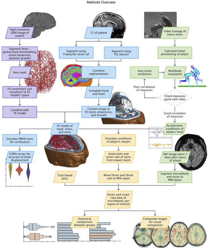
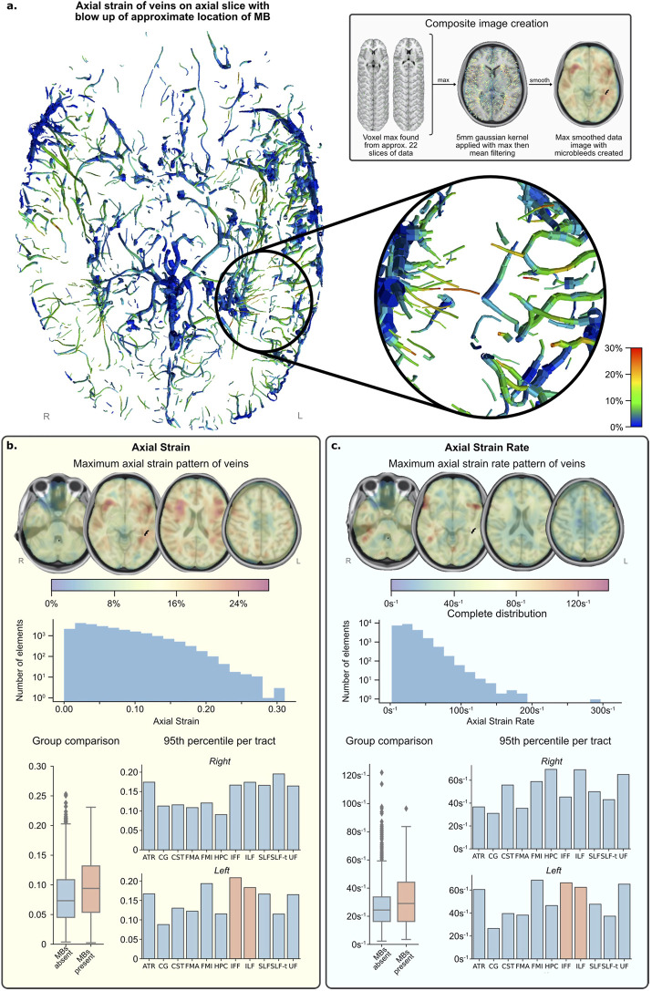
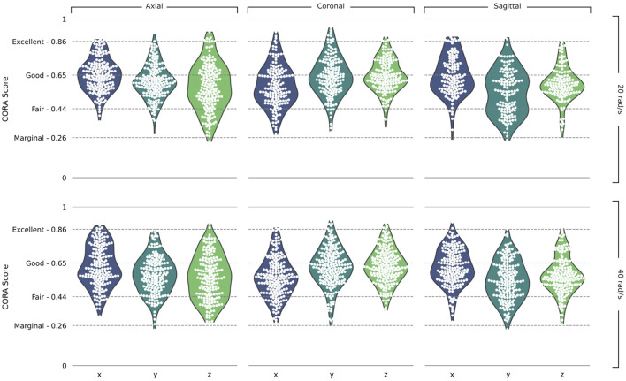
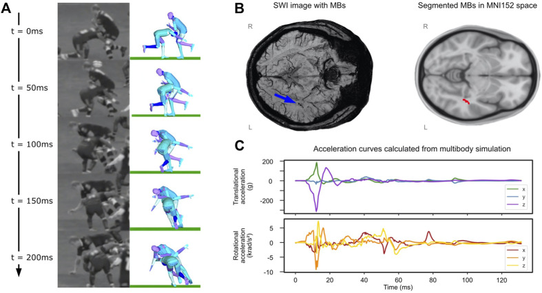
Finite Element (FE) models of brain mechanics have improved our understanding of the brain response to rapid mechanical loads that produce traumatic brain injuries. However, these models have rarely incorporated vasculature, which limits their ability to predict the response of vessels to head impacts. To address this shortcoming, here we used high-resolution MRI scans to map the venous system anatomy at a submillimetre resolution. We then used this map to develop an FE model of veins and incorporated it in an anatomically detailed FE model of the brain. The model prediction of brain displacement at different locations was compared to controlled experiments on post-mortem human subject heads, yielding over 3,100 displacement curve comparisons, which showed fair to excellent correlation between them. We then used the model to predict the distribution of axial strains and strain rates in the veins of a rugby player who had small blood deposits in his white matter, known as microbleeds, after sustaining a head collision. We hypothesised that the distribution of axial strain and strain rate in veins can predict the pattern of microbleeds. We reconstructed the head collision using video footage and multi-body dynamics modelling and used the predicted head accelerations to load the FE model of vascular injury. The model predicted large axial strains in veins where microbleeds were detected. A region of interest analysis using white matter tracts showed that the tract group with microbleeds had 95th percentile peak axial strain and strain rate of 0.197 and 64.9 s respectively, which were significantly larger than those of the group of tracts without microbleeds (0.163 and 57.0 s). This study does not derive a threshold for the onset of microbleeds as it investigated a single case, but it provides evidence for a link between strain and strain rate applied to veins during head impacts and structural damage and allows for future work to generate threshold values. Moreover, our results suggest that the FE model has the potential to be used to predict intracranial vascular injuries after TBI, providing a more objective tool for TBI assessment and improving protection against it.
脑力学的有限元(FE)模型增进了我们对大脑对导致创伤性脑损伤的快速机械负荷反应的理解。然而,这些模型很少纳入脉管系统,这限制了它们预测血管对头部撞击反应的能力。为了解决这一缺点,我们在此使用高分辨率MRI扫描以亚毫米分辨率绘制静脉系统解剖图。然后,我们利用这张图谱开发了一个静脉有限元模型,并将其纳入一个大脑解剖细节丰富的有限元模型中。将该模型对不同位置脑位移的预测与对尸体人类受试者头部进行的对照实验进行比较,得到了超过3100次位移曲线比较,结果显示二者之间具有良好到极佳的相关性。然后,我们使用该模型预测一名橄榄球运动员在头部碰撞后白质中出现小量血液沉积(即微出血)时其静脉内轴向应变和应变率的分布。我们假设静脉内轴向应变和应变率的分布可以预测微出血的模式。我们利用视频片段和多体动力学建模重建了头部碰撞过程,并使用预测的头部加速度对血管损伤有限元模型进行加载。该模型预测在检测到微出血的静脉处存在较大的轴向应变。使用白质束进行的感兴趣区域分析表明,有微出血的束组的第95百分位数峰值轴向应变和应变率分别为0.197和64.9/s,显著高于无微出血的束组(0.163和57.0/s)。本研究由于只调查了单个案例,并未得出微出血发生的阈值,但它为头部撞击期间施加于静脉的应变和应变率与结构损伤之间存在关联提供了证据,并为未来生成阈值的工作提供了可能。此外,我们的结果表明,有限元模型有潜力用于预测创伤性脑损伤后的颅内血管损伤,为创伤性脑损伤评估提供一个更客观的工具,并改善对其的防护。