Voltrova Barbora, Hybasek Vojtech, Blahnova Veronika, Sepitka Josef, Lukasova Vera, Vocetkova Karolina, Sovkova Vera, Matejka Roman, Fojt Jaroslav, Joska Ludek, Daniel Matej, Filova Eva
Department of Tissue Engineering, Institute of Experimental Medicine of the Czech Academy of Sciences Vídeňská 1083 142 20 Prague 4 Czech Republic
Charles University in Prague, Faculty of Science Albertov 2038/6 128 00 Prague Czech Republic.
RSC Adv. 2019 Apr 11;9(20):11341-11355. doi: 10.1039/c9ra00761j. eCollection 2019 Apr 9.
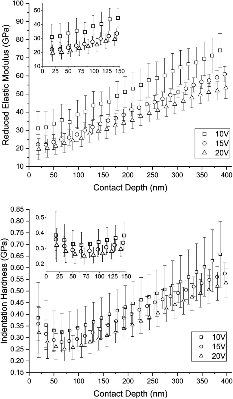
The formation of nanostructures on titanium implant surfaces is a promising strategy to modulate cell adhesion and differentiation, which are crucial for future application in bone regeneration. The aim of this study was to investigate how the nanotube diameter and/or nanomechanical properties alter human osteoblast like cell (Saos-2) adhesion, growth and osteogenic differentiation . Nanotubes, with diameters ranging from 24 to 66 nm, were fabricated on a commercially pure titanium (cpTi) surface using anodic oxidation with selected end potentials of 10 V, 15 V and 20 V. The cell response was studied on untreated and nanostructured samples using a measurement of metabolic activity, cell proliferation, alkaline phosphatase activity and qRT-PCR, which was used for the evaluation of osteogenic marker expression (collagen type I, osteocalcin, RunX2). Early cell adhesion was investigated using SEM and ELISA. Adhesive molecules (vinculin, talin), collagen and osteocalcin were also visualized using confocal microscopy. Moreover, the reduced elastic modulus and indentation hardness of nanotubes were assessed using a TriboIndenter™. Smooth and nanostructured cpTi both supported cell adhesion, proliferation and bone-specific mRNA expression. The nanotubes enhanced collagen type I and osteocalcin synthesis, compared to untreated cpTi, and the highest synthesis was observed on samples modified with 20 V nanotubes. Significant differences were found in the cell adhesion, where the vinculin and talin showed a dot-like distribution. Both the lowest reduced elastic modulus and indentation hardness were assessed from 20 V samples. The nanotubes of mainly 20 V samples showed a high potential for their use in bone implantation.
在钛植入物表面形成纳米结构是一种很有前景的策略,可用于调节细胞黏附与分化,这对其在骨再生中的未来应用至关重要。本研究的目的是探究纳米管直径和/或纳米力学性能如何改变人成骨样细胞(Saos-2)的黏附、生长和成骨分化。使用阳极氧化法,在商业纯钛(cpTi)表面制备了直径范围为24至66 nm的纳米管,选定的终电势为10 V、15 V和20 V。通过测量代谢活性、细胞增殖、碱性磷酸酶活性以及用于评估成骨标志物表达(I型胶原蛋白、骨钙素、RunX2)的qRT-PCR,对未处理和纳米结构化样品的细胞反应进行了研究。使用扫描电子显微镜(SEM)和酶联免疫吸附测定(ELISA)研究早期细胞黏附。还使用共聚焦显微镜观察了黏附分子(纽蛋白、踝蛋白)、胶原蛋白和骨钙素。此外,使用TriboIndenter™评估了纳米管的降低弹性模量和压痕硬度。光滑的和纳米结构化的cpTi均支持细胞黏附、增殖和骨特异性mRNA表达。与未处理的cpTi相比,纳米管增强了I型胶原蛋白和骨钙素的合成,在经20 V纳米管修饰的样品上观察到最高的合成量。在细胞黏附方面发现了显著差异,其中纽蛋白和踝蛋白呈点状分布。从20 V样品中评估出最低的降低弹性模量和压痕硬度。主要为20 V样品的纳米管在骨植入方面显示出很高的应用潜力。