Psychiatry Neuroimaging Laboratory, Department of Psychiatry, Brigham and Women's Hospital, Harvard Medical School, Boston, MA, USA.
Department of Psychiatry, Massachusetts General Hospital, Harvard Medical School, Boston, MA, USA.
Transl Psychiatry. 2022 May 7;12(1):191. doi: 10.1038/s41398-022-01960-8.
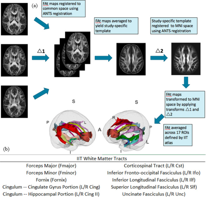
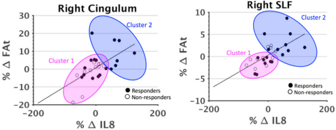
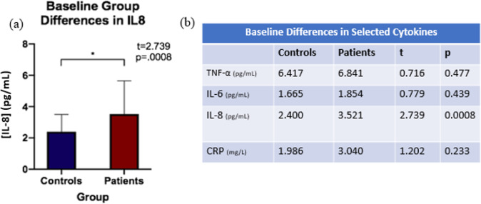
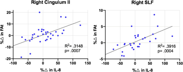
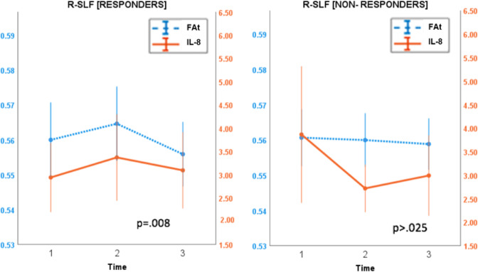
Research suggests electroconvulsive therapy (ECT) induces an acute neuroinflammatory response and changes in white matter (WM) structural connectivity. However, whether these processes are related, either to each other or to eventual treatment outcomes, has yet to be determined. We examined the relationship between levels of peripheral pro-inflammatory cytokines and diffusion imaging-indexed changes in WM microstructure in individuals with treatment-resistant depression (TRD) who underwent ECT. Forty-two patients were assessed at baseline, after their second ECT (T2), and after completion of ECT (T3). A Montgomery Åsberg Depression Rating Scale improvement of >50% post-ECT defined ECT-responders (n = 19) from non-responders (n = 23). Thirty-four controls were also examined. Tissue-specific fractional anisotropy (FAt) was estimated using diffusion imaging data and the Free-Water method in 17 WM tracts. Inflammatory panels were evaluated from peripheral blood. Cytokines were examined to characterize the association between potential ECT-induced changes in an inflammatory state and WM microstructure. Longitudinal trajectories of both measures were also examined separately for ECT-responders and non-responders. Patients exhibited elevated Interleukin-8 (IL-8) levels at baseline compared to controls. In patients, correlations between IL-8 and FAt changes from baseline to T2 were significant in the positive direction in the right superior longitudinal fasciculus (R-SLF) and right cingulum (R-CB) (p = 0.003). In these tracts, linear mixed-effects models revealed that trajectories of IL-8 and FAt were significantly positively correlated across all time points in responders, but not non-responders (R-CB-p = .001; R-SLF-p = 0.008). Our results suggest that response to ECT in TRD may be mediated by IL-8 and WM microstructure.
研究表明,电抽搐治疗(ECT)会引起急性神经炎症反应和白质(WM)结构连接的变化。然而,这些过程是否相互关联,或者与最终的治疗结果相关,尚未确定。我们研究了接受 ECT 的治疗抵抗性抑郁症(TRD)患者外周促炎细胞因子水平与 WM 微观结构扩散成像指标变化之间的关系。42 名患者在基线时、第二次 ECT 后(T2)和 ECT 完成后(T3)接受评估。ECT 后评分提高>50%定义为 ECT 反应者(n=19),评分未提高定义为非反应者(n=23)。还对 34 名对照进行了检查。使用扩散成像数据和 Free-Water 方法在 17 个 WM 束中估计组织特异性各向异性分数(FAt)。从外周血中评估炎症谱。检查细胞因子以表征炎症状态和 WM 微观结构之间潜在的 ECT 诱导变化之间的关联。还分别为 ECT 反应者和非反应者检查了这两种测量的纵向轨迹。与对照组相比,患者在基线时表现出较高的白细胞介素-8(IL-8)水平。在患者中,从基线到 T2 的 IL-8 和 FAt 变化之间的相关性在右侧上纵束(R-SLF)和右侧扣带束(R-CB)呈正相关(p=0.003)。在这些束中,线性混合效应模型显示,在反应者中,IL-8 和 FAt 的轨迹在所有时间点均呈显著正相关,但在非反应者中则不相关(R-CB-p=0.001;R-SLF-p=0.008)。我们的结果表明,TRD 中 ECT 的反应可能由 IL-8 和 WM 微观结构介导。