Al Ewaidat Haytham, Ayasrah Mohammad
Department of Allied Medical Sciences-Radiologic Technology, Faculty of Applied Medical Sciences, Jordan University of Science and Technology, Jordan.
Jordan University of Science and Technology, Department of Allied Medical Sciences-Radiologic Technology, Faculty of Applied Medical Sciences, Jordan University of Science and Technology, Jordan.
Int J Biomed Imaging. 2022 Apr 28;2022:8705531. doi: 10.1155/2022/8705531. eCollection 2022.
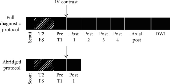
Breast MRI possesses high sensitivity for detecting breast cancer among the current clinical modalities and is an indispensable imaging practice. Breast MRI comprises diffusion-weighted imaging, ultrafast, and T2 weighted and T1 weighted CE (contrast-enhanced) imaging that may be utilized for improving the characterization of the lesions. This multimodal evaluation of breast lesions enables outstanding discrimination between the malignant and benign and malignant lesions. The expanding indications of breast MRI confirm the far superiority of MRI in preoperative staging, especially in the estimation of tumour size and identifying tumour foci in the contralateral and ipsilateral breast. Recent studies depicted that experts can meritoriously utilize this tool for improving breast cancer surgery despite their existence of no significant long term outcomes. For managing the, directly and indirectly, associated screening cost, abbreviated protocols are found to be more beneficial. Further, in some of the patients who were treated with neoadjuvant chemotherapy, breast MRI is utilized for documenting response. It is therefore essential to realise that oncological screening must be easily available, cost-effective, and time-consuming. Earlier detection of this short sequence protocol leads to prior and early breast cancer disease in high risky female populations like women with dense breasts, prehistoric evidence, etc. This proper utilization of AP reduces unnecessary mastectomies. Hence, this review focused on the explorative information for strongly suggesting the benefits of AP breast MRI compared to full diagnostic protocol MRI.
在目前的临床检查手段中,乳腺磁共振成像(MRI)对乳腺癌的检测具有高灵敏度,是一种不可或缺的影像学检查方法。乳腺MRI包括扩散加权成像、超快成像以及T2加权和T1加权对比增强成像,可用于提高病变特征的识别。这种对乳腺病变的多模态评估能够出色地区分恶性和良性病变。乳腺MRI适应证的不断扩大证实了其在术前分期方面的显著优势,尤其是在估计肿瘤大小以及识别对侧和同侧乳腺中的肿瘤病灶方面。最近的研究表明,尽管目前尚无显著的长期结果,但专家们能够出色地利用这一工具改善乳腺癌手术。为了直接或间接地控制相关的筛查成本,发现简化方案更具优势。此外,在一些接受新辅助化疗的患者中,乳腺MRI用于记录治疗反应。因此,必须认识到肿瘤筛查必须易于实施、具有成本效益且耗时较短。这种短序列方案的早期检测能够在高风险女性人群(如乳腺致密的女性、有家族病史的女性等)中更早地发现乳腺癌。这种适当使用乳腺MRI的方法可减少不必要的乳房切除术。因此,本综述着重探讨相关信息,以有力地表明与全诊断方案MRI相比,乳腺MRI简化方案的优势。