Ren Mindong, Xu Yang, Phoon Colin K L, Erdjument-Bromage Hediye, Neubert Thomas A, Rajan Sujith, Hussain M Mahmood, Schlame Michael
Departments of Anesthesiology, New York, NY, United States.
Department of Cell Biology, New York, NY, United States.
Front Cell Dev Biol. 2022 Apr 21;10:867175. doi: 10.3389/fcell.2022.867175. eCollection 2022.
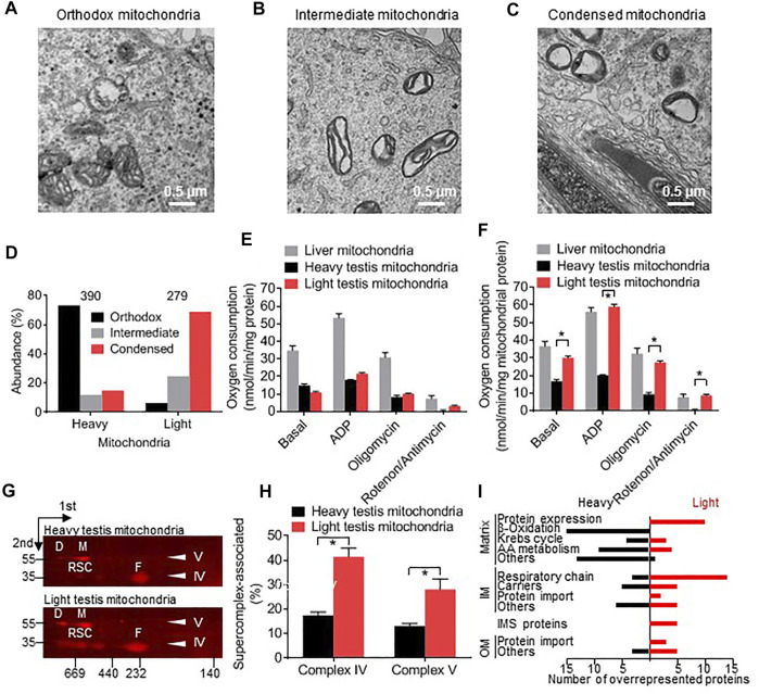
Mammalian spermatogenesis is associated with the transient appearance of condensed mitochondria, a singularity of germ cells with unknown function. Using proteomic analysis, respirometry, and electron microscopy with tomography, we studied the development of condensed mitochondria. Condensed mitochondria arose from orthodox mitochondria during meiosis by progressive contraction of the matrix space, which was accompanied by an initial expansion and a subsequent reduction of the surface area of the inner membrane. Compared to orthodox mitochondria, condensed mitochondria respired more actively, had a higher concentration of respiratory enzymes and supercomplexes, and contained more proteins involved in protein import and expression. After the completion of meiosis, the abundance of condensed mitochondria declined, which coincided with the onset of the biogenesis of acrosomes. Immuno-electron microscopy and the analysis of sub-cellular fractions suggested that condensed mitochondria or their fragments were translocated into the lumen of the acrosome. Thus, it seems condensed mitochondria are formed from orthodox mitochondria by extensive transformations in order to support the formation of the acrosomal matrix.
哺乳动物精子发生与凝聚线粒体的短暂出现有关,这是生殖细胞的一个独特现象,其功能尚不清楚。我们运用蛋白质组学分析、呼吸测定法以及电子显微镜断层扫描技术,研究了凝聚线粒体的发育过程。凝聚线粒体在减数分裂期间由正常线粒体通过基质空间的逐渐收缩形成,这一过程伴随着内膜表面积的先扩张后减少。与正常线粒体相比,凝聚线粒体呼吸更活跃,呼吸酶和超复合物浓度更高,且含有更多参与蛋白质导入和表达的蛋白质。减数分裂完成后,凝聚线粒体的丰度下降,这与顶体生物发生的开始相吻合。免疫电子显微镜和亚细胞组分分析表明,凝聚线粒体或其片段被转运到顶体腔内。因此,凝聚线粒体似乎是由正常线粒体通过广泛转变形成的,以支持顶体基质的形成。