Department of Physiology and Biomedical Sciences, Seoul National University College of Medicine, Seoul, 03080, South Korea.
Department of Internal Medicine, Seoul National University Hospital, Seoul, 03080, South Korea.
Commun Biol. 2022 May 9;5(1):431. doi: 10.1038/s42003-022-03388-8.
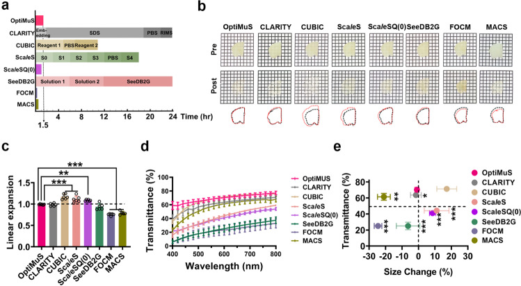
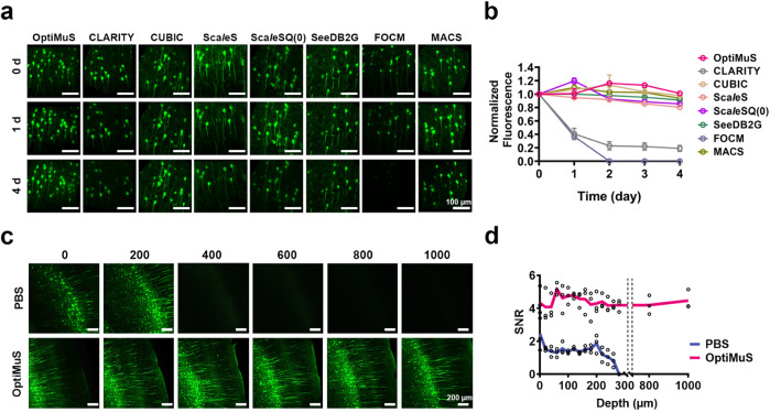
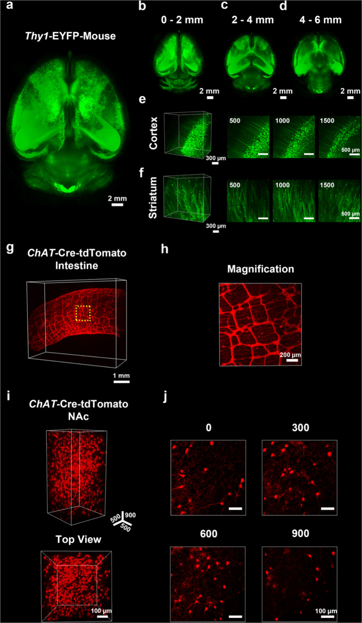
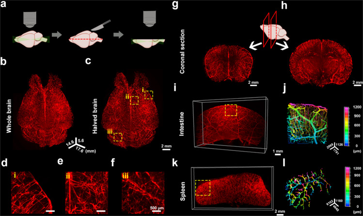
Various optical clearing approaches have been introduced to meet the growing demand for 3D volume imaging of biological structures. Each has its own strengths but still suffers from low transparency, long incubation time, processing complexity, tissue deformation, or fluorescence quenching, and a single solution that best satisfies all aspects has yet been developed. Here, we develop OptiMuS, an optimized single-step solution that overcomes the shortcomings of the existing aqueous-based clearing methods and that provides the best performance in terms of transparency, clearing rate, and size retention. OptiMuS achieves rapid and high transparency of brain tissues and other intact organs while preserving the size and fluorescent signal of the tissues. Moreover, OptiMuS is compatible with the use of lipophilic dyes, revealing DiI-labeled vascular structures of the whole brain, kidney, spleen, and intestine, and is also applied to 3D quantitative and comparative analysis of DiI-labeled vascular structures of glomeruli turfs in normal and diseased kidneys. Together, OptiMuS provides a single-step solution for simple, fast, and versatile optical clearing method to obtain high tissue transparency with minimum structural changes and is widely applicable for 3D imaging of various whole biological structures.
各种光学透明化方法已经被引入以满足对生物结构的 3D 体积成像的不断增长的需求。每种方法都有其自身的优势,但仍存在低透明度、长孵育时间、处理复杂性、组织变形或荧光淬灭的问题,并且尚未开发出一种能够满足所有方面的最佳单一解决方案。在这里,我们开发了 OptiMuS,这是一种优化的单步解决方案,克服了现有基于水的透明化方法的缺点,并在透明度、透明化速率和尺寸保留方面提供了最佳性能。OptiMuS 实现了脑组织和其他完整器官的快速高透明度,同时保持了组织的大小和荧光信号。此外,OptiMuS 与亲脂性染料兼容,可显示整个大脑、肾脏、脾脏和肠道的 DiI 标记的血管结构,还应用于正常和患病肾脏中 DiI 标记的肾小球草丛血管结构的 3D 定量和比较分析。总之,OptiMuS 为简单、快速和通用的光学透明化方法提供了一种单一的解决方案,以获得最小结构变化的高组织透明度,并且广泛适用于各种整个生物结构的 3D 成像。