Department of Radiological and Medical Laboratory Sciences, Nagoya University Graduate School of Medicine, Higashi-ku, Nagoya, Japan.
Department of Radiological Technology, Nagoya University Hospital, Showa-ku, Nagoya, Japan.
J Appl Clin Med Phys. 2022 Jul;23(7):e13626. doi: 10.1002/acm2.13626. Epub 2022 May 10.
Accurate tracer accumulation evaluation is difficult owing to the partial volume effect (PVE). We proposed a novel semi-quantitative approach for measuring the accumulation amount by examining the approximate image. Using a striatal phantom, we verified the validity of a newly proposed method to accurately evaluate the tracer accumulations in the caudate and putamen separately. Moreover, we compared the proposed method with the conventional methods.
The left and right caudate/putamen regions and the whole brain region as background were identified in computed tomography (CT) images obtained by single-photon emission computed tomography (SPECT)/CT and acquired the positional information of each region. SPECT-like images were generated by assigning assumed accumulation amounts to each region. The SPECT-like image, approximated to the actual measured SPECT image, was examined by changing the assumed accumulation amounts assigned to each region. When the generated SPECT-like image most approximated the actual measured SPECT image, the accumulation amounts assumed were determined as the accumulation amounts in each region. We evaluated the correlation between the count density calculated by the proposed method and the actual count density of the I solution filled in the phantom. Conventional methods (CT-guide method, geometric transfer matrix [GTM] method, region-based voxel-wise [RBV] method, and Southampton method) were also evaluated. The significance of differences between the correlation coefficients of various methods (except the Southampton method) was evaluated.
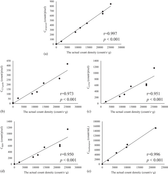
The correlation coefficients between the actual count density and the SPECT count densities were 0.997, 0.973, 0.951, 0.950, and 0.996 for the proposed method, CT-guide method, GTM method, RBV method, and Southampton method, respectively. The correlation of the proposed method was significantly higher than those of the other methods.
The proposed method could calculate accurate accumulation amounts in the caudate and putamen separately, considering the PVE.
由于部分容积效应(PVE),准确评估示踪剂的积累量较为困难。我们提出了一种新的半定量方法,通过检查近似图像来测量积累量。使用纹状体模型,我们验证了一种新方法来准确评估尾状核和壳核中示踪剂积累量的有效性。此外,我们将该方法与传统方法进行了比较。
在单光子发射计算机断层扫描(SPECT)/计算机断层扫描(CT)获得的 CT 图像中,识别左、右尾状核/壳核和整个大脑区域作为背景,并获取每个区域的位置信息。通过向每个区域分配假定的积累量来生成 SPECT 样图像。通过改变分配给每个区域的假定积累量,检查与实际测量的 SPECT 图像近似的 SPECT 样图像。当生成的 SPECT 样图像最接近实际测量的 SPECT 图像时,假定的积累量被确定为每个区域的积累量。我们评估了该方法计算的计数密度与在模型中填充的 I 溶液的实际计数密度之间的相关性。还评估了传统方法(CT 引导法、几何转移矩阵 [GTM] 法、基于区域的体素法 [RBV] 法和南安普顿法)。评估了各种方法(除了南安普顿法)的相关系数之间的差异的显著性。
对于该方法、CT 引导法、GTM 法、RBV 法和南安普顿法,实际计数密度与 SPECT 计数密度之间的相关系数分别为 0.997、0.973、0.951、0.950 和 0.996。该方法的相关性明显高于其他方法。
该方法考虑到 PVE,可以计算出尾状核和壳核中准确的积累量。