• 文献检索
  • 文档翻译
  • 深度研究
  • 学术资讯
  • Suppr Zotero 插件Zotero 插件
  • 邀请有礼
  • 套餐&价格
  • 历史记录
应用&插件
Suppr Zotero 插件Zotero 插件浏览器插件Mac 客户端Windows 客户端微信小程序
定价
高级版会员购买积分包购买API积分包
服务
文献检索文档翻译深度研究API 文档MCP 服务
关于我们
关于 Suppr公司介绍联系我们用户协议隐私条款
关注我们

Suppr 超能文献

核心技术专利:CN118964589B侵权必究
粤ICP备2023148730 号-1Suppr @ 2026

文献检索

告别复杂PubMed语法,用中文像聊天一样搜索,搜遍4000万医学文献。AI智能推荐,让科研检索更轻松。

立即免费搜索

文件翻译

保留排版,准确专业,支持PDF/Word/PPT等文件格式,支持 12+语言互译。

免费翻译文档

深度研究

AI帮你快速写综述,25分钟生成高质量综述,智能提取关键信息,辅助科研写作。

立即免费体验

磁性相互作用及生物相容性咖啡酸稳定的Fe-Pt簇作为MRI造影剂的研究

Magnetic interactions and study of biocompatible hydrocaffeic acid-stabilized Fe-Pt clusters as MRI contrast agents.

作者信息

Kostevšek N, Hudoklin S, Kreft M E, Serša I, Sepe A, Jagličić Z, Vidmar J, Ščančar J, Šturm S, Kobe S, Žužek Rožman K

机构信息

Department for Nanostructured Materials, Jožef Stefan Institute Jamova 39 Ljubljana Slovenia

Institute of Cell Biology, Faculty of Medicine, University of Ljubljana Vrazov trg 2 Ljubljana Slovenia.

出版信息

RSC Adv. 2018 Apr 19;8(26):14694-14704. doi: 10.1039/c8ra00047f. eCollection 2018 Apr 17.

DOI:10.1039/c8ra00047f
PMID:35540786
原文链接:https://pmc.ncbi.nlm.nih.gov/articles/PMC9080024/
Abstract

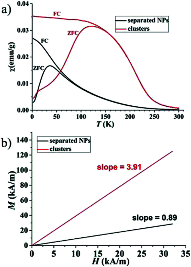
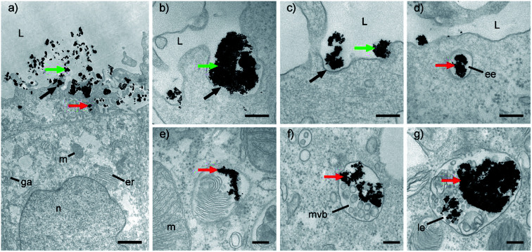
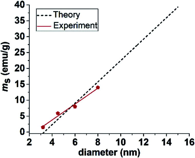
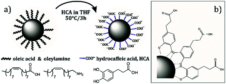
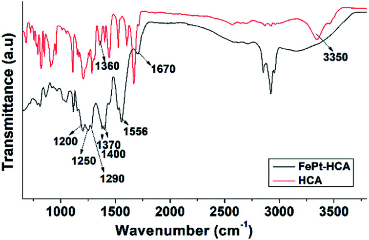
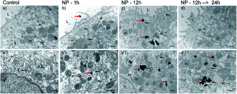
A detailed magnetic study of separated Fe-Pt NPs and Fe-Pt clusters was performed to predict their optimal size and morphology for the maximum saturation magnetization, a factor that is known to influence the performance of a magnetic-resonance-imaging (MRI) contrast agent. Excellent stability and biocompatibility of the nanoparticle suspension was achieved using a novel coating based on hydrocaffeic acid (HCA), which was confirmed with a detailed Fourier-transform infrared spectroscopy (FTIR) study. An study on a human-bladder papillary urothelial neoplasm RT4 cell line confirmed that HCA-Fe-Pt nanoparticles showed no cytotoxicity, even at a very high concentration (550 μg Fe-Pt per mL), with no delayed cytotoxic effect being detected. This indicates that the HCA coating provides excellent biocompatibility of the nanoparticles, which is a prerequisite for the material to be used as a safe contrast agent for MRI. The cellular uptake and internalization mechanism were studied using ICP-MS and TEM analyses. Furthermore, it was shown that even a very low concentration of Fe-Pt nanoparticles (<10 μg mL) in the cells is enough to decrease the relaxation times by 70%. In terms of the MRI imaging, this means a large improvement in the contrast, even at a low nanoparticle concentration and an easier visualization of the tissues containing nanoparticles, proving that HCA-coated Fe-Pt nanoparticles have the potential to be used as an efficient and safe MRI contrast agent.

摘要

对分离出的Fe-Pt纳米颗粒和Fe-Pt团簇进行了详细的磁性研究,以预测它们的最佳尺寸和形态,从而实现最大饱和磁化强度,这是一个已知会影响磁共振成像(MRI)造影剂性能的因素。使用基于氢咖啡酸(HCA)的新型涂层实现了纳米颗粒悬浮液的出色稳定性和生物相容性,这通过详细的傅里叶变换红外光谱(FTIR)研究得到了证实。对人膀胱乳头状尿路上皮肿瘤RT4细胞系的研究证实,即使在非常高的浓度(每毫升550μg Fe-Pt)下,HCA-Fe-Pt纳米颗粒也没有显示出细胞毒性,未检测到延迟的细胞毒性作用。这表明HCA涂层为纳米颗粒提供了出色的生物相容性,这是该材料用作安全的MRI造影剂的先决条件。使用电感耦合等离子体质谱(ICP-MS)和透射电子显微镜(TEM)分析研究了细胞摄取和内化机制。此外,研究表明,即使细胞中Fe-Pt纳米颗粒的浓度非常低(<10μg/mL),也足以使弛豫时间降低70%。就MRI成像而言,这意味着即使在低纳米颗粒浓度下对比度也有很大提高,并且更容易观察到含有纳米颗粒的组织,证明HCA包覆的Fe-Pt纳米颗粒有潜力用作高效、安全的MRI造影剂。

https://cdn.ncbi.nlm.nih.gov/pmc/blobs/b210/9080024/ab91318b369a/c8ra00047f-f6.jpg
https://cdn.ncbi.nlm.nih.gov/pmc/blobs/b210/9080024/514787b08d4b/c8ra00047f-f1.jpg
https://cdn.ncbi.nlm.nih.gov/pmc/blobs/b210/9080024/5344e377bfac/c8ra00047f-f2.jpg
https://cdn.ncbi.nlm.nih.gov/pmc/blobs/b210/9080024/0a2d15df7caa/c8ra00047f-f3.jpg
https://cdn.ncbi.nlm.nih.gov/pmc/blobs/b210/9080024/722faed8daf6/c8ra00047f-f4.jpg
https://cdn.ncbi.nlm.nih.gov/pmc/blobs/b210/9080024/a4058bf52fd4/c8ra00047f-f5.jpg
https://cdn.ncbi.nlm.nih.gov/pmc/blobs/b210/9080024/ab91318b369a/c8ra00047f-f6.jpg

相似文献

1
Magnetic interactions and study of biocompatible hydrocaffeic acid-stabilized Fe-Pt clusters as MRI contrast agents.磁性相互作用及生物相容性咖啡酸稳定的Fe-Pt簇作为MRI造影剂的研究
RSC Adv. 2018 Apr 19;8(26):14694-14704. doi: 10.1039/c8ra00047f. eCollection 2018 Apr 17.
2
Conjugation Magnetic PAEEP-PLLA Nanoparticles with Lactoferrin as a Specific Targeting MRI Contrast Agent for Detection of Brain Glioma in Rats.以乳铁蛋白为特异性靶向MRI造影剂的共轭磁性PAEEP-PLLA纳米颗粒用于大鼠脑胶质瘤检测
Nanoscale Res Lett. 2016 Dec;11(1):227. doi: 10.1186/s11671-016-1421-x. Epub 2016 Apr 27.
3
Uniform mesoporous silica coated iron oxide nanoparticles as a highly efficient, nontoxic MRI T(2) contrast agent with tunable proton relaxivities.均一介孔硅包覆氧化铁纳米颗粒作为一种高效、无毒的 MRI T(2) 造影剂,具有可调的质子弛豫率。
Contrast Media Mol Imaging. 2012 Sep-Oct;7(5):460-8. doi: 10.1002/cmmi.1473.
4
Water-soluble superparamagnetic magnetite nanoparticles with biocompatible coating for enhanced magnetic resonance imaging.具有生物相容性涂层的水溶性超顺磁磁铁矿纳米粒子,用于增强磁共振成像。
ACS Nano. 2011 Aug 23;5(8):6315-24. doi: 10.1021/nn201348s. Epub 2011 Aug 2.
5
Non-immunogenic dextran-coated superparamagnetic iron oxide nanoparticles: a biocompatible, size-tunable contrast agent for magnetic resonance imaging.非免疫原性葡聚糖包被的超顺磁性氧化铁纳米颗粒:一种用于磁共振成像的生物相容性、尺寸可调的造影剂。
Int J Nanomedicine. 2017 Jul 24;12:5223-5238. doi: 10.2147/IJN.S138108. eCollection 2017.
6
Trastuzumab conjugated porphyrin-superparamagnetic iron oxide nanoparticle: A potential PTT-MRI bimodal agent for herceptin positive breast cancer.曲妥珠单抗偶联卟啉-超顺磁性氧化铁纳米颗粒:一种用于赫赛汀阳性乳腺癌的潜在光热疗法-磁共振成像双模态剂。
Photodiagnosis Photodyn Ther. 2020 Sep;31:101896. doi: 10.1016/j.pdpdt.2020.101896. Epub 2020 Jun 22.
7
Synthesis of biocompatible, mesoporous Fe(3)O(4) nano/microspheres with large surface area for magnetic resonance imaging and therapeutic applications.合成具有大比表面积的生物相容性介孔 Fe(3)O(4)纳米/微球,用于磁共振成像和治疗应用。
ACS Appl Mater Interfaces. 2011 Feb;3(2):237-44. doi: 10.1021/am1012358. Epub 2011 Jan 13.
8
Suspension of Fe(3)O(4) nanoparticles stabilized by chitosan and o-carboxymethylchitosan.由壳聚糖和邻羧甲基壳聚糖稳定的Fe(3)O(4)纳米颗粒悬浮液。
Int J Pharm. 2008 Feb 28;350(1-2):361-8. doi: 10.1016/j.ijpharm.2007.09.004. Epub 2007 Sep 6.
9
Biocompatible Peptide-Coated Ultrasmall Superparamagnetic Iron Oxide Nanoparticles for In Vivo Contrast-Enhanced Magnetic Resonance Imaging.用于体内对比增强磁共振成像的生物相容型肽涂层超顺磁性氧化铁纳米颗粒。
ACS Nano. 2018 Jul 24;12(7):6480-6491. doi: 10.1021/acsnano.7b07572. Epub 2018 Jul 11.
10
Composites of aminodextran-coated Fe3O4 nanoparticles and graphene oxide for cellular magnetic resonance imaging.氨基葡聚糖包覆的 Fe3O4 纳米粒子与氧化石墨烯的复合材料用于细胞磁共振成像。
ACS Appl Mater Interfaces. 2011 Oct;3(10):4085-91. doi: 10.1021/am2009647. Epub 2011 Sep 19.

引用本文的文献

1
Nanogallium-poly(L-lactide) Composites with Contact Antibacterial Action.具有接触抗菌作用的纳米镓-聚(L-丙交酯)复合材料
Pharmaceutics. 2024 Feb 4;16(2):228. doi: 10.3390/pharmaceutics16020228.
2
Magneto-Liposomes as MRI Contrast Agents: A Systematic Study of Different Liposomal Formulations.磁性脂质体作为磁共振成像造影剂:不同脂质体制剂的系统研究
Nanomaterials (Basel). 2020 May 6;10(5):889. doi: 10.3390/nano10050889.

本文引用的文献

1
Increased endocytosis of magnetic nanoparticles into cancerous urothelial cells versus normal urothelial cells.与正常尿路上皮细胞相比,磁性纳米颗粒在癌性尿路上皮细胞中的内吞作用增强。
Histochem Cell Biol. 2018 Jan;149(1):45-59. doi: 10.1007/s00418-017-1605-1. Epub 2017 Aug 18.
2
Keeping it real: The importance of material characterization in nanotoxicology.实事求是:材料表征在纳米毒理学中的重要性。
Biochem Biophys Res Commun. 2015 Dec 18;468(3):498-503. doi: 10.1016/j.bbrc.2015.06.178. Epub 2015 Jul 15.
3
PEGylated FePt-Fe3O4 composite nanoassemblies (CNAs): in vitro hyperthermia, drug delivery and generation of reactive oxygen species (ROS).
聚乙二醇化的 FePt-Fe3O4 复合纳米组装体(CNAs):体外热疗、药物传递和活性氧(ROS)的产生。
Dalton Trans. 2015 May 21;44(19):9103-13. doi: 10.1039/c4dt03470h.
4
Water-soluble L-cysteine-coated FePt nanoparticles as dual MRI/CT imaging contrast agent for glioma.水溶性L-半胱氨酸包覆的FePt纳米颗粒作为用于胶质瘤的双模态MRI/CT成像造影剂
Int J Nanomedicine. 2015 Mar 23;10:2325-33. doi: 10.2147/IJN.S75174. eCollection 2015.
5
Facile surface functionalization of hydrophobic magnetic nanoparticles.疏水性磁性纳米颗粒的简便表面功能化
J Am Chem Soc. 2014 Sep 10;136(36):12552-5. doi: 10.1021/ja5060324. Epub 2014 Aug 28.
6
Uptake of fluorescent iron oxide nanoparticles by oligodendroglial OLN-93 cells.少突胶质细胞 OLN-93 细胞对荧光氧化铁纳米颗粒的摄取。
Neurochem Res. 2014 Feb;39(2):372-83. doi: 10.1007/s11064-013-1234-6. Epub 2013 Dec 25.
7
Gold nanoparticles as physiological markers of urine internalization into urothelial cells in vivo.金纳米颗粒作为尿液内吞进入体内尿路上皮细胞的生理标记物。
Int J Nanomedicine. 2013;8:3945-53. doi: 10.2147/IJN.S44363. Epub 2013 Oct 14.
8
Assessing nanoparticle toxicity in cell-based assays: influence of cell culture parameters and optimized models for bridging the in vitro-in vivo gap.基于细胞的分析评估纳米颗粒的毒性:细胞培养参数的影响和优化模型,以弥合体外-体内差距。
Chem Soc Rev. 2013 Nov 7;42(21):8339-59. doi: 10.1039/c3cs60145e. Epub 2013 Jul 23.
9
PEGylated FePt@Fe2O3 core-shell magnetic nanoparticles: potential theranostic applications and in vivo toxicity studies.聚乙二醇化 FePt@Fe2O3 核壳磁性纳米粒子:潜在的治疗应用和体内毒性研究。
Nanomedicine. 2013 Oct;9(7):1077-88. doi: 10.1016/j.nano.2013.02.010. Epub 2013 Mar 8.
10
Cooperative organization in iron oxide multi-core nanoparticles potentiates their efficiency as heating mediators and MRI contrast agents.铁氧化物多核纳米粒子的协同组织增强了它们作为热疗介质和 MRI 对比剂的效率。
ACS Nano. 2012 Dec 21;6(12):10935-49. doi: 10.1021/nn304477s. Epub 2012 Nov 29.