Liang Shuyan, Zhou Qing, Wang Min, Zhu Yanhong, Wu Qingzhi, Yang Xiangliang
College of Life Science and Technology, Huazhong University of Science and Technology, Wuhan, People's Republic of China.
State Key Laboratory of Advanced Technology for Materials Synthesis and Processing, Wuhan University of Technology, Wuhan, People's Republic of China ; Biomedical Material and Engineering Center of Hubei Province, Wuhan University of Technology, Wuhan, People's Republic of China.
Int J Nanomedicine. 2015 Mar 23;10:2325-33. doi: 10.2147/IJN.S75174. eCollection 2015.
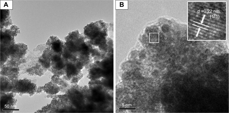
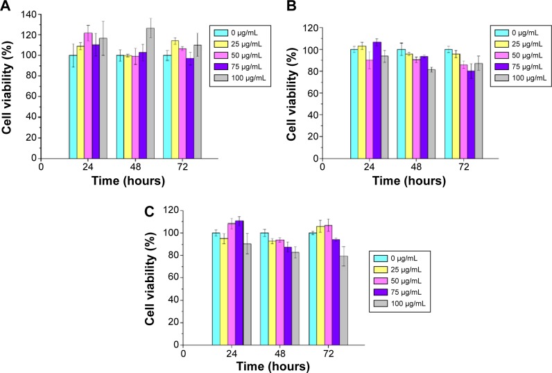
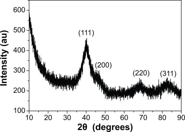
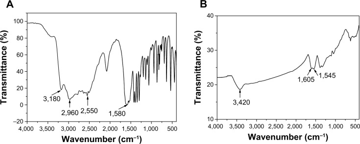
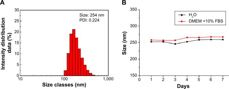
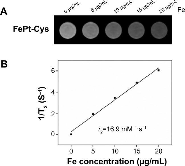
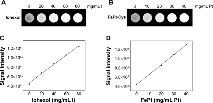
Nanoparticles (NPs) are advantageous for the delivery of diagnosis agents to brain tumors. In this study, we attempted to develop an L-cysteine coated FePt (FePt-Cys) NP as MRI/CT imaging contrast agent for the diagnosis of malignant gliomas. FePt-Cys NPs were synthesized through a co-reduction route, which was characterized by transmission electron microscopy, high-resolution transmission electron microscopy, powder X-ray diffraction, Fourier transform infrared spectroscopy, and dynamic light scattering. The MRI and CT imaging ability of FePt-Cys NPs was evaluated using different gliomas cells (C6, SGH44, U251) as the model. Furthermore, the biocompatibility of the as-synthesized FePt-Cys NPs was evaluated using three different cell lines (ECV304, L929, and HEK293) as the model. The results showed that FePt-Cys NPs displayed excellent biocompatibility and good MRI/CT imaging ability, thereby indicating promising potential as a dual MRI/CT contrast agent for the diagnosis of brain malignant gliomas.
纳米颗粒(NPs)在将诊断剂递送至脑肿瘤方面具有优势。在本研究中,我们试图开发一种L-半胱氨酸包覆的FePt(FePt-Cys)纳米颗粒作为磁共振成像(MRI)/计算机断层扫描(CT)成像造影剂用于恶性胶质瘤的诊断。FePt-Cys纳米颗粒通过共还原途径合成,并用透射电子显微镜、高分辨率透射电子显微镜、粉末X射线衍射、傅里叶变换红外光谱和动态光散射对其进行了表征。以不同的胶质瘤细胞(C6、SGH44、U251)为模型评估了FePt-Cys纳米颗粒的MRI和CT成像能力。此外,以三种不同的细胞系(ECV304、L929和HEK293)为模型评估了合成的FePt-Cys纳米颗粒的生物相容性。结果表明,FePt-Cys纳米颗粒表现出优异的生物相容性和良好的MRI/CT成像能力,从而表明其作为用于诊断脑恶性胶质瘤的双功能MRI/CT造影剂具有广阔的应用前景。