Biology Department, Duke University, Durham, NC 27708.
Department of Pharmacology and.
Mol Biol Cell. 2022 Sep 15;33(11):ar94. doi: 10.1091/mbc.E21-11-0537. Epub 2022 May 11.
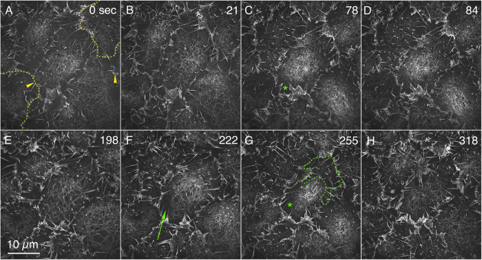
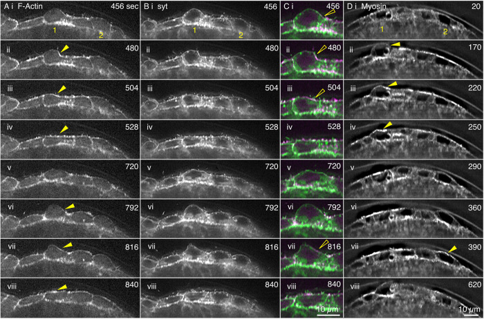
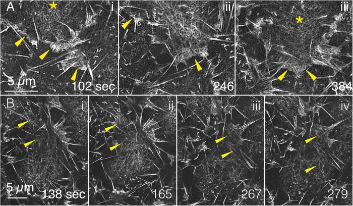
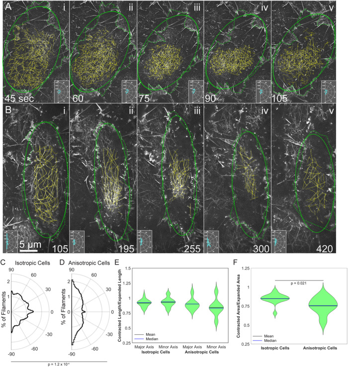
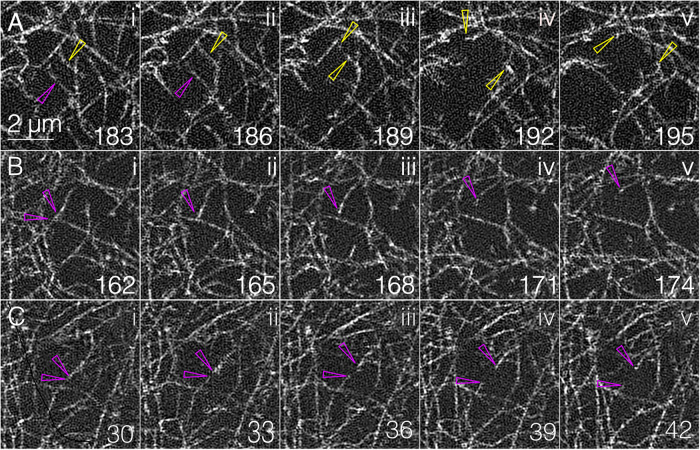
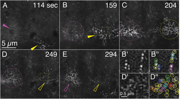
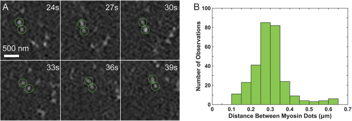
Arrays of actin filaments (F-actin) near the apical surface of epithelial cells (medioapical arrays) contribute to apical constriction and morphogenesis throughout phylogeny. Here, superresolution approaches (grazing incidence structured illumination, GI-SIM, and lattice light sheet, LLSM) microscopy resolve individual, fluorescently labeled F-actin and bipolar myosin filaments that drive amnioserosa cell shape changes during dorsal closure in . In expanded cells, F-actin and myosin form loose, apically domed meshworks at the plasma membrane. The arrays condense as cells contract, drawing the domes into the plane of the junctional belts. As condensation continues, individual filaments are no longer uniformly apparent. As cells expand, arrays of actomyosin are again resolved-some F-actin turnover likely occurs, but a large fraction of existing filaments rearrange. In morphologically isotropic cells, actin filaments are randomly oriented and during contraction are drawn together but remain essentially randomly oriented. In anisotropic cells, largely parallel actin filaments are drawn closer to one another. Our images offer unparalleled resolution of F-actin in embryonic tissue, show that medioapical arrays are tightly apposed to the plasma membrane and are continuous with meshworks of lamellar F-actin. Medioapical arrays thereby constitute modified cell cortex. In concert with other tagged array components, superresolution imaging of live specimens will offer new understanding of cortical architecture and function.
上皮细胞顶表面附近的肌动蛋白丝阵列(中顶阵列)有助于整个系统发育过程中的顶端收缩和形态发生。在这里,超分辨率方法(掠入射结构照明、GI-SIM 和晶格光片、LLSM)显微镜解析了单个荧光标记的 F-肌动蛋白和双极肌球蛋白丝,这些丝在 背腹闭合过程中驱动羊膜细胞形状变化。在扩展的细胞中,F-肌动蛋白和肌球蛋白在质膜上形成松散的、顶向拱形的网格。随着细胞收缩,阵列浓缩,将穹顶拉到连接带的平面上。随着浓缩的继续,单个丝不再均匀出现。随着细胞的扩展,肌动球蛋白阵列再次被解析-可能发生一些 F-肌动蛋白的周转,但很大一部分现有丝重新排列。在形态各向同性的细胞中,肌动蛋白丝随机取向,在收缩时被拉到一起,但基本保持随机取向。在各向异性的细胞中,大量平行的肌动蛋白丝彼此更靠近。我们的图像提供了胚胎组织中 F-肌动蛋白的无与伦比的分辨率,表明中顶阵列紧密贴合质膜,并与层状 F-肌动蛋白的网格连续。因此,中顶阵列构成了改良的细胞皮层。与其他标记的阵列组件一起,活标本的超分辨率成像将提供对皮质结构和功能的新理解。