Queiroz Diego, Lustosa Gustavo Porto, Mascato Diego, Morales Melina Correia, Fernandes Arthur Gustavo, Fernandes Rodrigo Antonio Brant
Ophthal Hospital Especializado Ltda, São Paulo, SP, Brasil.
Departamento de Oftalmologia e Ciências Visuais, Escola Paulista de Medicina, Universidade Federal de São Paulo, São Paulo, SP, Brasil.
Arq Bras Oftalmol. 2022 May 9;86(6). doi: 10.5935/0004-2749.20230073.
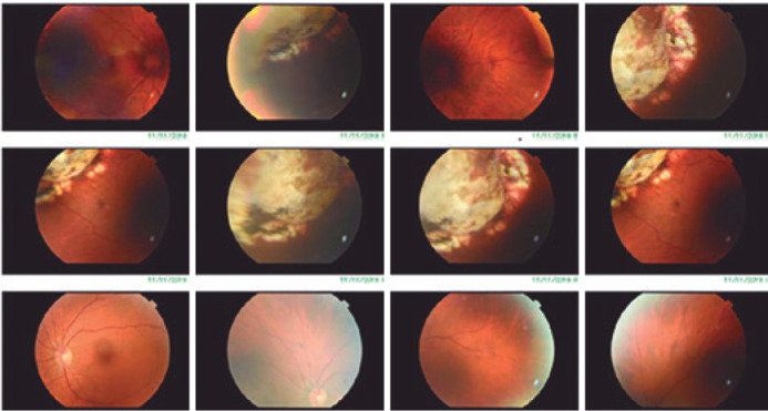
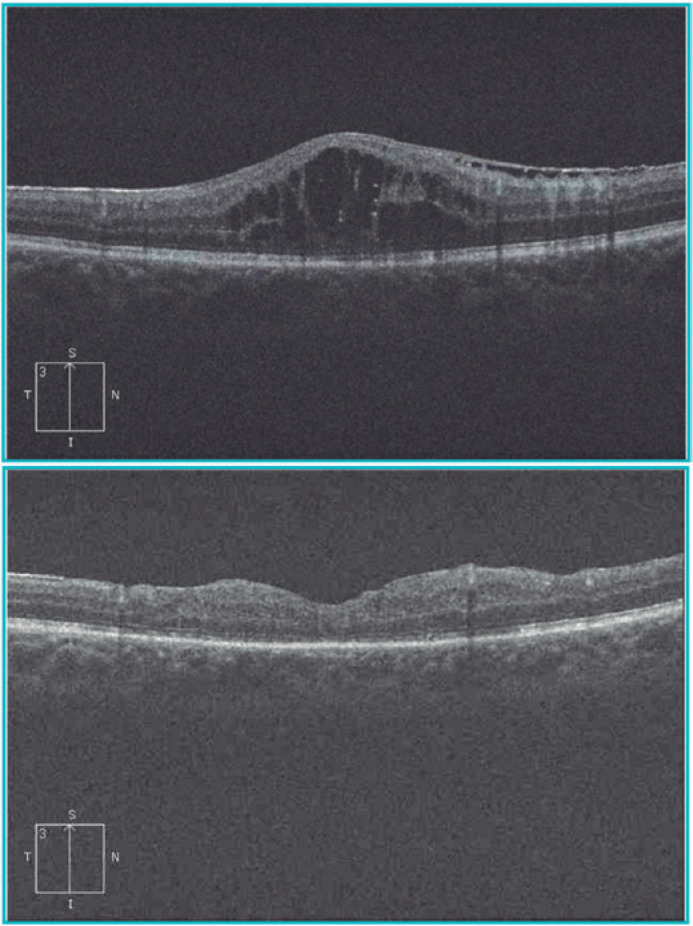
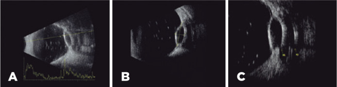
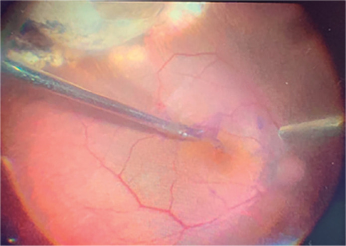
This is a case report involving a 56-year-old male patient with a history of pars plana vitrectomy due to a rhegmatogenous retinal detachment in the right eye that resulted in the implantation of a drainage device after the patient developed secondary glaucoma. Two years after the device's implantation, the patient was referred to our care as his visual acuity had decreased to 20/200 (1.00 LogMAR). At the fundus evaluation, a choroidal amelanotic elevation was observed at the upper temporal equator, and a potential diagnosis was made of amelanotic choroidal melanoma. The ultrasound exam visualized the patient's implanted superotemporal justabulbar drainage device, which revealed a transscleral communication from the plate fibrocapsular's draining space to the suprachoroidal space (fistula). The ultrasound also revealed a focal pocket of choroidal detachment in the patient's superotemporal region, simulating an amelanotic choroidal melanoma. A new pars plana vitrectomy was performed to remove the internal limiting membrane without repercussions at the fistula site. The patient's recovery progressed well, and he regained a visual acuity of 20/70 (0.55 LogMAR). To the best of our knowledge, this is the first case report of this condition.
这是一例病例报告,患者为一名56岁男性,有右眼孔源性视网膜脱离行玻璃体切除术病史,术后继发青光眼并植入了引流装置。该装置植入两年后,患者因视力下降至20/200(1.00 LogMAR)前来我院就诊。眼底检查时,在颞上赤道部观察到脉络膜无黑色素性隆起,初步诊断为无黑色素性脉络膜黑色素瘤。超声检查显示患者颞上球旁植入的引流装置,发现从板纤维囊引流间隙到脉络膜上腔存在经巩膜通道(瘘管)。超声还显示患者颞上区域有局限性脉络膜脱离区,类似无黑色素性脉络膜黑色素瘤。遂行再次玻璃体切除术以切除内界膜,瘘管部位未受影响。患者恢复良好,视力恢复至20/70(0.55 LogMAR)。据我们所知,这是该病症的首例病例报告。