• 文献检索
  • 文档翻译
  • 深度研究
  • 学术资讯
  • Suppr Zotero 插件Zotero 插件
  • 邀请有礼
  • 套餐&价格
  • 历史记录
应用&插件
Suppr Zotero 插件Zotero 插件浏览器插件Mac 客户端Windows 客户端微信小程序
定价
高级版会员购买积分包购买API积分包
服务
文献检索文档翻译深度研究API 文档MCP 服务
关于我们
关于 Suppr公司介绍联系我们用户协议隐私条款
关注我们

Suppr 超能文献

核心技术专利:CN118964589B侵权必究
粤ICP备2023148730 号-1Suppr @ 2026

文献检索

告别复杂PubMed语法,用中文像聊天一样搜索,搜遍4000万医学文献。AI智能推荐,让科研检索更轻松。

立即免费搜索

文件翻译

保留排版,准确专业,支持PDF/Word/PPT等文件格式,支持 12+语言互译。

免费翻译文档

深度研究

AI帮你快速写综述,25分钟生成高质量综述,智能提取关键信息,辅助科研写作。

立即免费体验

电子显微镜在肾小球疾病诊断与研究中的最新进展

Recent advances in electron microscopy for the diagnosis and research of glomerular diseases.

作者信息

Honda Kazuho, Takaki Takashi, Kang Dedong

机构信息

Department of Anatomy, Showa University School of Medicine, Tokyo, Japan.

Division of Electron Microscopy, Showa University, Tokyo, Japan.

出版信息

Kidney Res Clin Pract. 2023 Mar;42(2):155-165. doi: 10.23876/j.krcp.21.270. Epub 2022 May 4.

DOI:10.23876/j.krcp.21.270
PMID:35545227
原文链接:https://pmc.ncbi.nlm.nih.gov/articles/PMC10085717/
Abstract

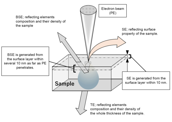
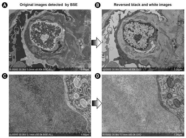
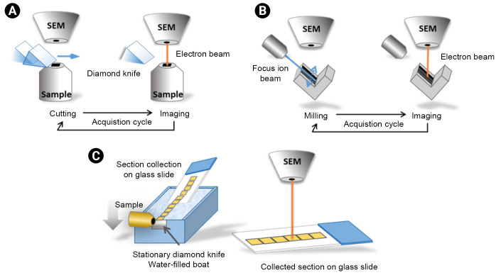
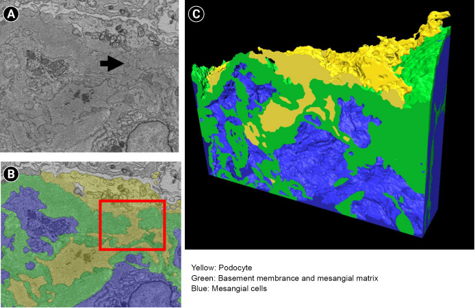
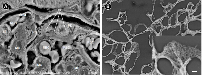
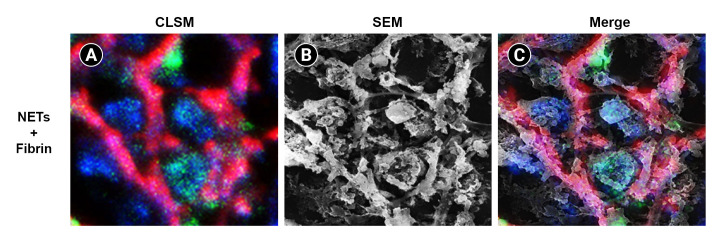
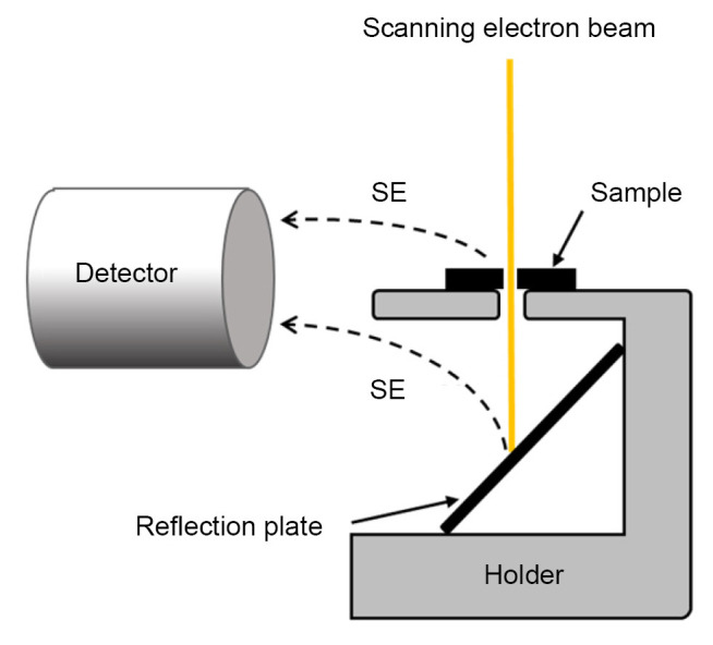
Recent technical advances in the detection of backscattered electrons during scanning electron microscopy (SEM) have improved resolution and have provided several new technologies for research and clinical practice in kidney disease. The advances include three-dimensional (3D) electron microscopy (3D-EM), correlative light and electron microscopy (CLEM), low-vacuum SEM (LVSEM), and scanning transmission electron microscopy (STEM). 3D-EM analysis used to be laborious, but recently three different technologies, serial block-face SEM, focused ion beam SEM, and array tomography, have made 3D-EM easier by automating sectioning and the subsequent image acquisition in an SEM. CLEM is a method to correlate light microscopic images, especially immunofluorescent and electron microscopy images, providing detailed ultrastructure of the area of interest where the immunofluorescent marker is located. LVSEM enables the use of SEM on materials with poor electron conductivity. For example, LVSEM makes it possible for high resolution, 3D observation of paraffin sections. Finally, STEM is a method to observe ultrathin sections with improved resolution by using the focused electron beam scanning used in SEM and not the broad electron beam used in transmission electron microscopy. These technical advances in electron microscopy are promising to provide plenty of novel insights for understanding the pathogenesis and diagnosis of various glomerular diseases.

摘要

扫描电子显微镜(SEM)中背散射电子检测技术的最新进展提高了分辨率,并为肾脏疾病的研究和临床实践提供了多种新技术。这些进展包括三维(3D)电子显微镜(3D-EM)、 correlative light and electron microscopy(CLEM)、低真空SEM(LVSEM)和扫描透射电子显微镜(STEM)。3D-EM分析过去很费力,但最近三种不同的技术,即连续块面SEM、聚焦离子束SEM和阵列断层扫描,通过在SEM中自动切片和随后的图像采集,使3D-EM变得更容易。CLEM是一种将光学显微镜图像,特别是免疫荧光和电子显微镜图像相关联的方法,可提供免疫荧光标记所在感兴趣区域的详细超微结构。LVSEM能够对电子导电性差的材料进行SEM分析。例如,LVSEM使对石蜡切片进行高分辨率、三维观察成为可能。最后,STEM是一种通过使用SEM中使用的聚焦电子束扫描而不是透射电子显微镜中使用的宽电子束来观察超薄切片的方法,具有更高的分辨率。电子显微镜的这些技术进展有望为理解各种肾小球疾病的发病机制和诊断提供大量新见解。

https://cdn.ncbi.nlm.nih.gov/pmc/blobs/3042/10085717/93737a61f63c/j-krcp-21-270f7.jpg
https://cdn.ncbi.nlm.nih.gov/pmc/blobs/3042/10085717/b610bb818092/j-krcp-21-270f1.jpg
https://cdn.ncbi.nlm.nih.gov/pmc/blobs/3042/10085717/49d4175488cb/j-krcp-21-270f2.jpg
https://cdn.ncbi.nlm.nih.gov/pmc/blobs/3042/10085717/d2c7e0ef0eb1/j-krcp-21-270f3.jpg
https://cdn.ncbi.nlm.nih.gov/pmc/blobs/3042/10085717/b94d21b75dbb/j-krcp-21-270f4.jpg
https://cdn.ncbi.nlm.nih.gov/pmc/blobs/3042/10085717/8a626d768256/j-krcp-21-270f5.jpg
https://cdn.ncbi.nlm.nih.gov/pmc/blobs/3042/10085717/9e83e31f5572/j-krcp-21-270f6.jpg
https://cdn.ncbi.nlm.nih.gov/pmc/blobs/3042/10085717/93737a61f63c/j-krcp-21-270f7.jpg

相似文献

1
Recent advances in electron microscopy for the diagnosis and research of glomerular diseases.电子显微镜在肾小球疾病诊断与研究中的最新进展
Kidney Res Clin Pract. 2023 Mar;42(2):155-165. doi: 10.23876/j.krcp.21.270. Epub 2022 May 4.
2
A workflow for 3D-CLEM investigating liver tissue.用于研究肝组织的 3D-CLEM 工作流程。
J Microsc. 2021 Mar;281(3):231-242. doi: 10.1111/jmi.12967. Epub 2020 Oct 27.
3
Applications of Scanning Electron Microscopy Using Secondary and Backscattered Electron Signals in Neural Structure.利用二次电子和背散射电子信号的扫描电子显微镜在神经结构中的应用
Front Neuroanat. 2021 Dec 2;15:759804. doi: 10.3389/fnana.2021.759804. eCollection 2021.
4
Combining serial block face and focused ion beam scanning electron microscopy for 3D studies of rare events.结合连续块面和聚焦离子束扫描电子显微镜用于罕见事件的三维研究。
Methods Cell Biol. 2019;152:87-101. doi: 10.1016/bs.mcb.2019.03.014. Epub 2019 May 7.
5
Reactive oxygen FIB spin milling enables correlative workflow for 3D super-resolution light microscopy and serial FIB/SEM of cultured cells.活性氧纤维铣削实现了用于培养细胞的 3D 超分辨率光显微镜和连续纤维切割/扫描电镜的相关工作流程。
Sci Rep. 2021 Jun 23;11(1):13162. doi: 10.1038/s41598-021-92608-y.
6
Backscattered electron imaging of resin-embedded sections.树脂包埋切片的背散射电子成像。
Microscopy (Oxf). 2018 Jun 15. doi: 10.1093/jmicro/dfy028.
7
Developing 3D SEM in a broad biological context.在广泛的生物学背景下开发三维扫描电子显微镜。
J Microsc. 2015 Aug;259(2):80-96. doi: 10.1111/jmi.12211. Epub 2015 Jan 26.
8
Advanced cryo-tomography workflow developments - correlative microscopy, milling automation and cryo-lift-out.先进的冷冻电子断层扫描工作流程发展——相关显微镜技术、铣削自动化和冷冻提取技术。
J Microsc. 2021 Feb;281(2):112-124. doi: 10.1111/jmi.12939. Epub 2020 Jul 2.
9
High-Resolution 3D Imaging of Megakaryocytes Using Focused Ion Beam-Scanning Electron Microscopy.使用聚焦离子束扫描电子显微镜对巨核细胞进行高分辨率三维成像
Methods Mol Biol. 2018;1812:217-231. doi: 10.1007/978-1-4939-8585-2_13.
10
The application and development of electron microscopy for three-dimensional reconstruction in life science: a review.电子显微镜在生命科学中三维重构的应用与发展:综述。
Cell Tissue Res. 2024 Apr;396(1):1-18. doi: 10.1007/s00441-024-03878-7. Epub 2024 Feb 28.

引用本文的文献

1
Fast Processing of Electron Microscopic Specimen Preserved Ultrastructure of Glomeruli and Electron-Dense Deposits in Diagnostic Renal Biopsies: A Prospective and Retrospective Comparative Study.快速处理诊断性肾活检中保存肾小球超微结构和电子致密沉积物的电子显微镜标本:一项前瞻性和回顾性比较研究。
Mod Pathol. 2025 Feb;38(2):100662. doi: 10.1016/j.modpat.2024.100662. Epub 2024 Nov 20.
2
Nanoscale Optical Imaging, Reconstruction, and Spatial Analysis of Whole Mouse Glomeruli.全小鼠肾小球的纳米级光学成像、重建与空间分析
bioRxiv. 2024 Dec 9:2024.10.31.620364. doi: 10.1101/2024.10.31.620364.
3
Serial Block Face-Scanning Electron Microscopy as a Burgeoning Technology.

本文引用的文献

1
Pathological Analysis of Early Transplant Glomerulopathy in Renal Allografts Using Low-Vacuum Scanning Electron Microscopy.低真空扫描电子显微镜在肾移植早期移植性肾小球病中的病理分析。
Nephron. 2020;144 Suppl 1(Suppl 1):71-78. doi: 10.1159/000512136. Epub 2020 Dec 1.
2
Three-dimensional electron microscopy for endothelial glycocalyx observation using Alcian blue with silver enhancement.使用阿利新蓝结合银增强技术的三维电子显微镜观察内皮糖萼。
Med Mol Morphol. 2021 Jun;54(2):95-107. doi: 10.1007/s00795-020-00267-1. Epub 2020 Oct 6.
3
Glomerular Cellular Interactions Following Disruption of the Glomerular Basement Membrane in IgA Nephropathy: Ultrastructural Analyses by 3-Dimensional Serial Block-Face Scanning Electron Microscopy.
序列块面扫描电子显微镜技术的兴起。
Adv Biol (Weinh). 2023 Aug;7(8):e2300139. doi: 10.1002/adbi.202300139. Epub 2023 May 28.
4
Updates in renal pathology.肾脏病理学的进展
Kidney Res Clin Pract. 2023 Mar;42(2):153-154. doi: 10.23876/j.krcp.22.299. Epub 2023 Mar 31.
IgA肾病中肾小球基底膜破坏后的肾小球细胞相互作用:三维连续块面扫描电子显微镜超微结构分析
Kidney Med. 2020 Feb 1;2(2):222-225. doi: 10.1016/j.xkme.2019.11.003. eCollection 2020 Mar-Apr.
4
Low-vacuum scanning electron microscopy may allow early diagnosis of human renal transplant antibody-mediated rejection.低真空扫描电子显微镜可能有助于早期诊断人类肾移植抗体介导的排斥反应。
Biomed Res. 2020;41(2):81-90. doi: 10.2220/biomedres.41.81.
5
Three-dimensional imaging of podocyte ultrastructure using FE-SEM and FIB-SEM tomography.使用 FE-SEM 和 FIB-SEM 断层扫描技术对足细胞超微结构进行三维成像。
Cell Tissue Res. 2020 Feb;379(2):245-254. doi: 10.1007/s00441-019-03118-3. Epub 2019 Nov 22.
6
Podocyte penetration of the glomerular basement membrane to contact on the mesangial cell at the lesion of mesangial interposition in lupus nephritis: a three-dimensional analysis by serial block-face scanning electron microscopy.狼疮性肾炎系膜插入病变时足细胞穿过肾小球基底膜与系膜细胞接触:连续块面扫描电子显微镜的三维分析。
Clin Exp Nephrol. 2019 Jun;23(6):773-781. doi: 10.1007/s10157-019-01701-0. Epub 2019 Feb 8.
7
Morphological Processes of Foot Process Effacement in Puromycin Aminonucleoside Nephrosis Revealed by FIB/SEM Tomography.FIB/SEM 断层扫描揭示嘌呤霉素氨基核苷肾病中足细胞足突消失的形态发生过程。
J Am Soc Nephrol. 2019 Jan;30(1):96-108. doi: 10.1681/ASN.2018020139. Epub 2018 Dec 4.
8
Large Volume Electron Microscopy and Neural Microcircuit Analysis.大体积电子显微镜与神经微回路分析。
Front Neural Circuits. 2018 Nov 12;12:98. doi: 10.3389/fncir.2018.00098. eCollection 2018.
9
Backscattered electron imaging of resin-embedded sections.树脂包埋切片的背散射电子成像。
Microscopy (Oxf). 2018 Jun 15. doi: 10.1093/jmicro/dfy028.
10
Informative three-dimensional survey of cell/tissue architectures in thick paraffin sections by simple low-vacuum scanning electron microscopy.简单低真空扫描电子显微镜对厚石蜡切片中细胞/组织结构的信息三维观察。
Sci Rep. 2018 May 10;8(1):7479. doi: 10.1038/s41598-018-25840-8.