• 文献检索
  • 文档翻译
  • 深度研究
  • 学术资讯
  • Suppr Zotero 插件Zotero 插件
  • 邀请有礼
  • 套餐&价格
  • 历史记录
应用&插件
Suppr Zotero 插件Zotero 插件浏览器插件Mac 客户端Windows 客户端微信小程序
定价
高级版会员购买积分包购买API积分包
服务
文献检索文档翻译深度研究API 文档MCP 服务
关于我们
关于 Suppr公司介绍联系我们用户协议隐私条款
关注我们

Suppr 超能文献

核心技术专利:CN118964589B侵权必究
粤ICP备2023148730 号-1Suppr @ 2026

文献检索

告别复杂PubMed语法,用中文像聊天一样搜索,搜遍4000万医学文献。AI智能推荐,让科研检索更轻松。

立即免费搜索

文件翻译

保留排版,准确专业,支持PDF/Word/PPT等文件格式,支持 12+语言互译。

免费翻译文档

深度研究

AI帮你快速写综述,25分钟生成高质量综述,智能提取关键信息,辅助科研写作。

立即免费体验

FIB/SEM 断层扫描揭示嘌呤霉素氨基核苷肾病中足细胞足突消失的形态发生过程。

Morphological Processes of Foot Process Effacement in Puromycin Aminonucleoside Nephrosis Revealed by FIB/SEM Tomography.

机构信息

Department of Anatomy and Life Structure and

Laboratory of Morphology and Image Analysis, Research Support Center, Juntendo University Graduate School of Medicine, Tokyo, Japan.

出版信息

J Am Soc Nephrol. 2019 Jan;30(1):96-108. doi: 10.1681/ASN.2018020139. Epub 2018 Dec 4.

DOI:10.1681/ASN.2018020139
PMID:30514724
原文链接:https://pmc.ncbi.nlm.nih.gov/articles/PMC6317610/
Abstract

BACKGROUND

Foot process effacement is one of the pathologic indicators of podocyte injury. However, the morphologic changes associated with it remain unclear.

METHODS

To clarify the developmental process, we analyzed puromycin nephrotic podocytes reconstructed from serial focused-ion beam/scanning electron microscopy (FIB/SEM) images.

RESULTS

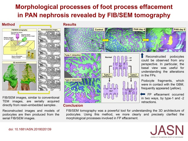
Intact podocytes consisted of four subcellular compartments: cell body, primary process, ridge-like prominence (RLP), and foot process. The RLP, a longitudinal protrusion from the basal surface of the cell body and primary process, served as an adhesive apparatus for the cell body and primary process to attach to the glomerular basement membrane. Foot processes protruded from both sides of the RLP. In puromycin nephrotic podocytes, foot process effacement occurred in two ways: by type-1 retraction, where the foot processes retracted while maintaining their rounded tips; or type-2 retraction, where they narrowed across their entire lengths, tapering toward the tips. Puromycin nephrotic podocytes also exhibited several alterations associated with foot process effacement, such as deformation of the cell body, retraction of RLPs, and cytoplasmic fragmentation. Finally, podocytes were reorganized into a broad, flattened shape.

CONCLUSIONS

The three-dimensional reconstruction of podocytes by serial FIB/SEM images revealed the morphologic changes involved in foot process effacement in greater detail than previously described.

摘要

背景

足细胞裂孔消失是足细胞损伤的病理指标之一。然而,与之相关的形态变化仍不清楚。

方法

为了阐明这一过程,我们对从小鼠足细胞裂孔消失模型中分离得到的、经连续聚焦离子束/扫描电镜(FIB/SEM)成像重构的足细胞进行了分析。

结果

完整的足细胞由四个亚细胞区室组成:细胞体、初级突起、嵴样隆起(RLP)和足突。RLP 是从细胞体和初级突起的基底面纵向伸出的突起,是将细胞体和初级突起附着到肾小球基底膜的附着装置。足突从 RLP 的两侧伸出。在嘌呤霉素肾病足细胞中,足突裂孔消失有两种方式:1 型回缩,足突回缩但保持圆顶状;2 型回缩,整个足突变窄,向顶端逐渐变细。嘌呤霉素肾病足细胞还表现出与足突裂孔消失相关的几种改变,如细胞体变形、RLP 回缩和细胞质碎片化。最后,足细胞被重新组织成宽阔的扁平形状。

结论

通过连续 FIB/SEM 图像对足细胞进行三维重建,比以前的描述更详细地揭示了足突裂孔消失所涉及的形态变化。

https://cdn.ncbi.nlm.nih.gov/pmc/blobs/a783/6317610/2646c2195eaa/ASN.2018020139absf1.jpg

相似文献

1
Morphological Processes of Foot Process Effacement in Puromycin Aminonucleoside Nephrosis Revealed by FIB/SEM Tomography.FIB/SEM 断层扫描揭示嘌呤霉素氨基核苷肾病中足细胞足突消失的形态发生过程。
J Am Soc Nephrol. 2019 Jan;30(1):96-108. doi: 10.1681/ASN.2018020139. Epub 2018 Dec 4.
2
Re-evaluation of foot process effacement in acute puromycin aminonucleoside nephrosis.急性嘌呤霉素氨基核苷肾病足突消失的重新评估
Kidney Int. 1996 Oct;50(4):1278-87. doi: 10.1038/ki.1996.439.
3
Three-dimensional imaging of podocyte ultrastructure using FE-SEM and FIB-SEM tomography.使用 FE-SEM 和 FIB-SEM 断层扫描技术对足细胞超微结构进行三维成像。
Cell Tissue Res. 2020 Feb;379(2):245-254. doi: 10.1007/s00441-019-03118-3. Epub 2019 Nov 22.
4
Ultrastructural changes in glomerular epithelial cells in acute puromycin aminonucleoside nephrosis: a study by high-resolution scanning electron microscopy.急性嘌呤霉素氨基核苷肾病肾小球上皮细胞的超微结构变化:高分辨率扫描电子显微镜研究
Virchows Arch A Pathol Anat Histopathol. 1993;423(2):111-9. doi: 10.1007/BF01606585.
5
Tolvaptan, a selective oral vasopressin V2 receptor antagonist, ameliorates podocyte injury in puromycin aminonucleoside nephrotic rats.托伐普坦,一种选择性口服血管加压素V2受体拮抗剂,可改善嘌呤霉素氨基核苷肾病大鼠的足细胞损伤。
Clin Exp Nephrol. 2009 Oct;13(5):438-446. doi: 10.1007/s10157-009-0196-0. Epub 2009 May 19.
6
Podocin is translocated to cytoplasm in puromycin aminonucleoside nephrosis rats and in poor-prognosis patients with IgA nephropathy.在嘌呤霉素氨基核苷肾病大鼠和预后不良的IgA肾病患者中,足突蛋白转位至细胞质。
Cell Tissue Res. 2015 May;360(2):391-400. doi: 10.1007/s00441-014-2100-9. Epub 2015 Feb 13.
7
Triptolide protects podocytes from puromycin aminonucleoside induced injury in vivo and in vitro.雷公藤甲素在体内和体外均可保护足细胞免受嘌呤霉素氨基核苷诱导的损伤。
Kidney Int. 2008 Sep;74(5):596-612. doi: 10.1038/ki.2008.203. Epub 2008 May 28.
8
Upregulation of nestin protects podocytes from apoptosis induced by puromycin aminonucleoside.巢蛋白的上调可保护足细胞免受嘌呤霉素氨基核苷诱导的细胞凋亡。
Am J Nephrol. 2011;34(5):423-34. doi: 10.1159/000331701. Epub 2011 Sep 21.
9
Role of Fat1 in cell-cell contact formation of podocytes in puromycin aminonucleoside nephrosis and neonatal kidney.Fat1在嘌呤霉素氨基核苷肾病和新生肾脏足细胞细胞间接触形成中的作用。
Kidney Int. 2005 Aug;68(2):542-51. doi: 10.1111/j.1523-1755.2005.00432.x.
10
The relationship between glomerular function and podocyte structure of pre-proteinuria and acute nephrosis in puromycin aminonucleoside-induced rat models: a comparative electron microscopic study.嘌呤霉素氨基核苷诱导大鼠模型中蛋白尿前期和急性肾病的肾小球功能与足细胞结构的关系:一项对比电子显微镜研究
Rom J Morphol Embryol. 2017;58(3):823-830.

引用本文的文献

1
Primary cilium disappearance in podocytes during vertebrate phylogeny revealed by array tomography.阵列断层扫描揭示脊椎动物系统发育过程中足细胞初级纤毛的消失
Cell Tissue Res. 2025 Aug 29. doi: 10.1007/s00441-025-04002-z.
2
Advancing the clinical assessment of glomerular podocyte pathology in kidney biopsies via super-resolution microscopy and angiopoietin-like 4 staining.通过超分辨率显微镜和血管生成素样4染色推进肾活检中肾小球足细胞病理的临床评估。
Theranostics. 2025 Jan 1;15(3):784-803. doi: 10.7150/thno.101498. eCollection 2025.
3
mTOR pathway: A key player in diabetic nephropathy progression and therapeutic targets.

本文引用的文献

1
Backscattered electron imaging of resin-embedded sections.树脂包埋切片的背散射电子成像。
Microscopy (Oxf). 2018 Jun 15. doi: 10.1093/jmicro/dfy028.
2
A Device for Ribbon Collection for Array Tomography with Scanning Electron Microscopy.一种用于扫描电子显微镜阵列断层成像的带状收集装置。
Acta Histochem Cytochem. 2017 Oct 24;50(5):135-140. doi: 10.1267/ahc.17013. Epub 2017 Oct 19.
3
Morphological process of podocyte development revealed by block-face scanning electron microscopy.通过块面扫描电子显微镜揭示的足细胞发育的形态学过程。
mTOR信号通路:糖尿病肾病进展中的关键因素及治疗靶点。
Genes Dis. 2024 Mar 8;12(2):101260. doi: 10.1016/j.gendis.2024.101260. eCollection 2025 Mar.
4
ATP dynamics as a predictor of future podocyte structure and function after acute ischemic kidney injury in female mice.ATP 动态变化可预测雌性小鼠急性缺血性肾损伤后足细胞结构和功能的未来变化。
Nat Commun. 2024 Nov 22;15(1):9977. doi: 10.1038/s41467-024-54222-0.
5
Paraoxonase 2 (PON2) Deficiency Reproduces Lipid Alterations of Diabetic and Inflammatory Glomerular Disease and Affects TRPC6 Signaling.对氧磷酶 2 (PON2) 缺乏可重现糖尿病和炎症性肾小球疾病的脂质改变,并影响 TRPC6 信号转导。
Cells. 2022 Nov 16;11(22):3625. doi: 10.3390/cells11223625.
6
Recent advances in electron microscopy for the diagnosis and research of glomerular diseases.电子显微镜在肾小球疾病诊断与研究中的最新进展
Kidney Res Clin Pract. 2023 Mar;42(2):155-165. doi: 10.23876/j.krcp.21.270. Epub 2022 May 4.
7
Quantitative Analyses of Foot Processes, Mitochondria, and Basement Membranes by Structured Illumination Microscopy Using Elastica-Masson- and Periodic-Acid-Schiff-Stained Kidney Sections.使用弹性纤维-马松染色法和过碘酸-希夫染色法对肾脏切片进行结构照明显微镜检查,对足突、线粒体和基底膜进行定量分析。
Kidney Int Rep. 2021 May 1;6(7):1923-1938. doi: 10.1016/j.ekir.2021.04.021. eCollection 2021 Jul.
8
Super-resolved local recruitment of CLDN5 to filtration slits implicates a direct relationship with podocyte foot process effacement.超分辨局部募集 Claudin5 到滤过裂隙,暗示其与足细胞足突消失有直接关系。
J Cell Mol Med. 2021 Aug;25(16):7631-7641. doi: 10.1111/jcmm.16519. Epub 2021 Jun 22.
9
Therapeutic trials in adult FSGS: lessons learned and the road forward.成人 FSGS 的治疗试验:经验教训与前进道路。
Nat Rev Nephrol. 2021 Sep;17(9):619-630. doi: 10.1038/s41581-021-00427-1. Epub 2021 May 20.
10
Three-Dimensional Architecture of Glomerular Endothelial Cells Revealed by FIB-SEM Tomography.聚焦离子束扫描电子显微镜断层扫描揭示肾小球内皮细胞的三维结构
Front Cell Dev Biol. 2021 Mar 11;9:653472. doi: 10.3389/fcell.2021.653472. eCollection 2021.
J Cell Sci. 2017 Jan 1;130(1):132-142. doi: 10.1242/jcs.187815. Epub 2016 Jun 29.
4
Evolutionary morphology of podocytes and primary urine-producing apparatus.足细胞与原尿生成装置的进化形态学
Anat Sci Int. 2017 Mar;92(2):161-172. doi: 10.1007/s12565-015-0317-7. Epub 2015 Dec 1.
5
Advanced electron microscopic techniques provide a deeper insight into the peculiar features of podocytes.先进的电子显微镜技术能更深入地洞察足细胞的独特特征。
Am J Physiol Renal Physiol. 2015 Dec 15;309(12):F1082-9. doi: 10.1152/ajprenal.00338.2015. Epub 2015 Sep 23.
6
Three-dimensional architecture of podocytes revealed by block-face scanning electron microscopy.通过块面扫描电子显微镜揭示的足细胞三维结构
Sci Rep. 2015 Mar 11;5:8993. doi: 10.1038/srep08993.
7
Podocytes … What's Under Yours? (Podocytes and Foot Processes and How They Change in Nephropathy).足细胞……你的下面是什么?(足细胞和足突以及它们在肾病中的变化)。
Front Endocrinol (Lausanne). 2015 Feb 23;6:9. doi: 10.3389/fendo.2015.00009. eCollection 2015.
8
Structural analysis of how podocytes detach from the glomerular basement membrane under hypertrophic stress.足细胞在肥大应激下如何从肾小球基底膜脱离的结构分析。
Front Endocrinol (Lausanne). 2014 Dec 12;5:207. doi: 10.3389/fendo.2014.00207. eCollection 2014.
9
A potential role for mechanical forces in the detachment of podocytes and the progression of CKD.机械力在足细胞脱离及慢性肾脏病进展中的潜在作用。
J Am Soc Nephrol. 2015 Feb;26(2):258-69. doi: 10.1681/ASN.2014030278. Epub 2014 Jul 24.
10
Array tomography.面分选术
J Microsc. 2013 Nov;252(2):93-9. doi: 10.1111/jmi.12087. Epub 2013 Sep 20.