Department of Neurosurgery, University Medical Center Mainz, Langenbeckstr. 1, 55131, Mainz, Germany.
Department of Neurosurgery, Klinik Hirslanden, Zurich, Switzerland.
Neurosurg Rev. 2022 Aug;45(4):2887-2894. doi: 10.1007/s10143-022-01794-4. Epub 2022 May 12.
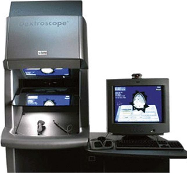
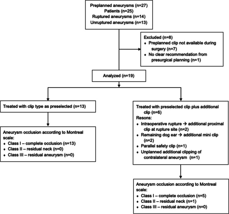
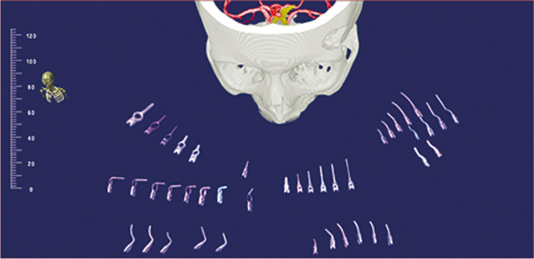
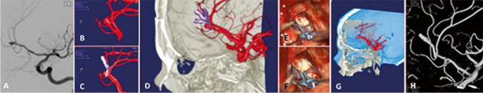
Aneurysm occlusion rate after clipping is higher than after endovascular treatment. However, a certain percentage of incompletely clipped aneurysms remains. Presurgical selection of the proper aneurysm clips could potentially reduce the rate of incomplete clippings caused by inadequate clip geometry. The aim of the present study was to assess whether preoperative 3D image-based simulation allows for preoperative selection of a proper aneurysm clip for complete occlusion in individual cases. Patients harboring ruptured or unruptured cerebral aneurysms prior to surgical clipping were analyzed. CT angiography images were transferred to a 3D surgical-planning station (Dextroscope®) with imported models of 58 aneurysm clips. Intracranial vessels and aneurysms were segmented and the virtual aneurysm clips were placed at the aneurysm neck. Operating surgeons had information about the selected aneurysm clip, and patients underwent clipping. Intraoperative clip selection was documented and aneurysm occlusion rate was assessed by postoperative digital subtraction angiography. Nineteen patients were available for final analysis. In all patients, the most proximal clip at the aneurysm neck was the preselected clip. All aneurysms except one were fully occluded, as assessed by catheter angiography. One aneurysm had a small neck remnant that did not require secondary surgery and was occluded 15 months after surgery. 3D image-based preselection of a proper aneurysm clip can be translated to the operating room and avoids intraoperative clip selection. The associated occlusion rate of aneurysms is high.
夹闭术后的动脉瘤闭塞率高于血管内治疗。然而,仍有一定比例的未完全夹闭的动脉瘤。术前选择合适的动脉瘤夹可能会降低因夹合几何形状不合适而导致不完全夹闭的发生率。本研究的目的是评估术前基于 3D 图像的模拟是否可以在个体病例中选择合适的动脉瘤夹以实现完全闭塞。分析了术前接受夹闭手术的破裂或未破裂脑动脉瘤患者。将 CT 血管造影图像传输到带有 58 种动脉瘤夹导入模型的 3D 手术规划工作站(Dextroscope®)。颅内血管和动脉瘤被分割,虚拟动脉瘤夹被放置在动脉瘤颈部。手术医生获得关于所选动脉瘤夹的信息,患者接受夹闭手术。记录术中夹的选择,并通过术后数字减影血管造影评估动脉瘤闭塞率。19 例患者可进行最终分析。在所有患者中,动脉瘤颈部最接近的夹闭夹是预先选择的夹闭夹。所有动脉瘤均完全闭塞(根据导管血管造影评估),除 1 个动脉瘤外。1 个动脉瘤颈残留小,不需要二次手术,术后 15 个月闭塞。基于 3D 图像的合适动脉瘤夹预选可以转化到手术室,并避免术中夹选择。相关的动脉瘤闭塞率较高。