Kong Ming, Xu Manman, Zhou Ying, Geng Nan, Lin Ning, Song Wenyan, Li Shanshan, Piao Yuetong, Han Zuoqing, Guo Rong, Yang Chao, Luo Nan, Wang Zhong, Ma Lei, Xu Quanxiao, Wang Lili, Qiu Wanchun, Li Junfeng, Shi Daimeng, Cheung Eddie C, Li Rongkuan, Chen Yu, Duan Zhongping
Beijing Municipal Key Laboratory of Liver Failure and Artificial Liver Treatment Research, Fourth Department of Liver Disease, Beijing Youan Hospital, Capital Medical University, Beijing, China.
Postgraduate Training Base of Jinzhou Medical University, Department of Gastroenterology and Hepatology, The Chinese People's Liberation Army Rocket Force Characteristic Medical Center, Beijing, China.
Front Nutr. 2022 Apr 25;9:871697. doi: 10.3389/fnut.2022.871697. eCollection 2022.
Abdominal adipose is closely related to many endocrine and metabolic diseases. The aim of this study was to analyze the distribution of abdominal adipose tissue in a healthy population in northern China determined by abdominal computed tomography (CT).
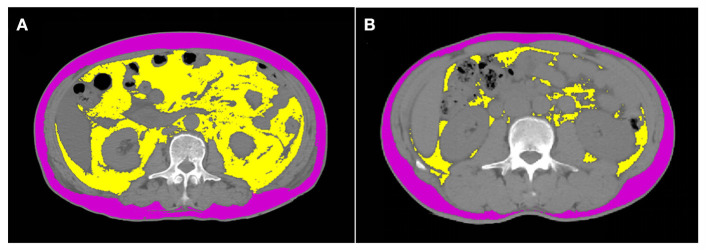
Data for this study were obtained from a multicenter, retrospective, cross-sectional study that collected abdominal CT scans of 1787 healthy individuals from 4 representative cities in northern China. Areas of visceral adipose tissue (VATA) and subcutaneous adipose tissue (SATA) were obtained by measuring CT images at the level of the 3rd lumbar vertebra. Visceral adipose tissue index (VATI) and subcutaneous adipose index (SATI) were obtained by normalizing the square of height to analyze the distribution of the above indexes and visceral obesity among different body mass index (BMI), gender and age.
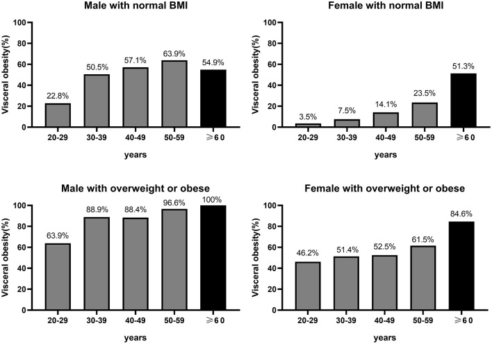
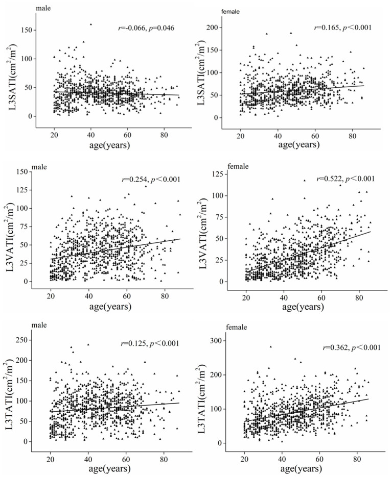
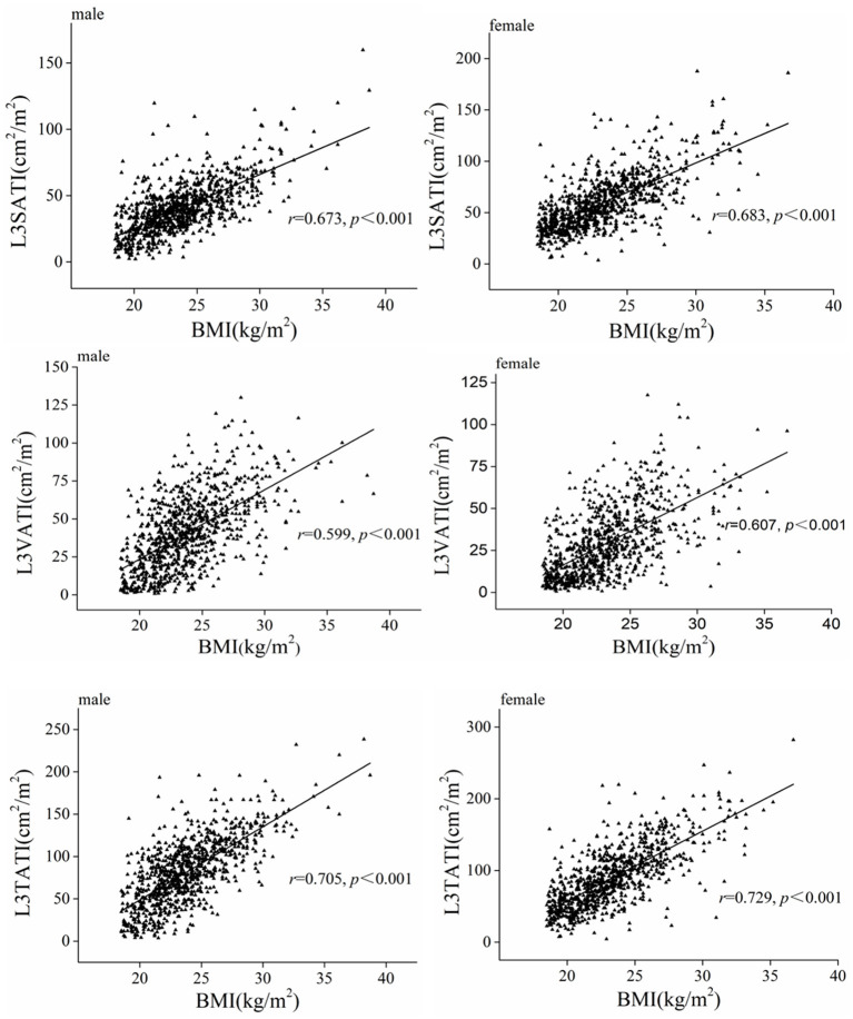
The mean age of this healthy population was 45.3 ± 15.2 years and the mean BMI was 23.5 ± 3.2 kg/m, with 902 men and 885 women. Compared with women, men had a significantly higher median VATA (120.9 vs. 67.2 cm), VATI (39.1 vs. 25.6 cm/m) and a significantly higher percentage of visceral adiposity (VATA ≥ 100 cm) (60.8 vs. 30.4%), while women had significantly higher SATA (116.9 vs. 146.7 cm) and SATI (38.8 vs. 55.8 cm/m) than men. Whether men or women, VATI was positively correlated with age. Interestingly, SATI was weakly positively correlated with age in women, while SATI was weakly negatively correlated with age in men. In persons with a normal BMI, the proportion of visceral adiposity increases with age, whereas in men with a normal BMI, the proportion of visceral adiposity decreases after the age of 60 years but remains >50%.
The distribution of abdominal visceral and subcutaneous adipose tissue parameters measured by CT differed among gender, age, and BMI. Even men and women with normal BMI have a high proportion of visceral obesity.
腹部脂肪与多种内分泌和代谢疾病密切相关。本研究旨在分析通过腹部计算机断层扫描(CT)确定的中国北方健康人群腹部脂肪组织的分布情况。
本研究数据来自一项多中心、回顾性横断面研究,收集了中国北方4个代表性城市1787名健康个体的腹部CT扫描图像。通过测量第3腰椎水平的CT图像获得内脏脂肪组织(VATA)和皮下脂肪组织(SATA)的面积。通过将身高平方标准化来获得内脏脂肪组织指数(VATI)和皮下脂肪指数(SATI),以分析上述指标及内脏肥胖在不同体重指数(BMI)、性别和年龄中的分布情况。
该健康人群的平均年龄为45.3±15.2岁,平均BMI为23.5±3.2kg/m²,其中男性902人,女性885人。与女性相比,男性的VATA中位数(120.9对67.2cm²)、VATI(39.1对25.6cm²/m²)显著更高,内脏肥胖百分比(VATA≥100cm²)也显著更高(60.8%对30.4%),而女性的SATA(116.9对146.7cm²)和SATI(38.8对55.8cm²/m²)显著高于男性。无论男性还是女性,VATI均与年龄呈正相关。有趣的是,女性中SATI与年龄呈弱正相关,而男性中SATI与年龄呈弱负相关。在BMI正常的人群中,内脏肥胖比例随年龄增加而增加,而在BMI正常的男性中,60岁后内脏肥胖比例下降,但仍>50%。
通过CT测量的腹部内脏和皮下脂肪组织参数的分布在性别、年龄和BMI之间存在差异。即使BMI正常的男性和女性也有较高比例的内脏肥胖。