Service de Radiologie, APHP Hôpitaux Paris Saclay, Hôpital Antoine Béclère, 157 rue de la porte de trivaux, 92140, Clamart, France.
Service de Radiopédiatrie, Armand Trousseau Hospital, Paris, France.
Eur J Med Res. 2022 May 12;27(1):67. doi: 10.1186/s40001-022-00692-1.
We report the challenging case of a 6-year-old boy with precocious puberty related to histologically proven Leydig cell tumor.
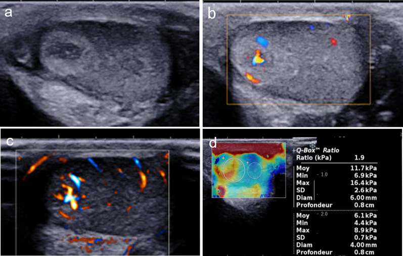
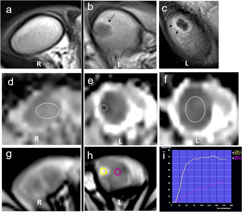
Multiparametric ultrasound and magnetic resonance imaging (MRI) was performed. Interesting findings were scarcely or never reported in children and differed from adults Leydig cell tumors s such as the hyperechogenic halo surrounding the lesion and the dominant central vascularization using ultrasensitive Doppler. MRI revealed an enlarged testicle with strong enhancement of a tumor, a tumor apparent diffusion coefficient (ADC) of 600 × 10 mm/s and a lower ADC value of the non-tumor parenchyma compared to the contralateral testis (ADC = 800 × 10 mm/s vs 1100 × 10 mm/s), attributed to the spermatogenesis induced by hormonal impregnation.
We illustrate multiparametric US and MRI findings of a pediatric Leydig cell tumor, including the imaging changes attributed to local hormone secretion, which may be helpful in similar cases.
我们报告了一例 6 岁男孩早发性性早熟的罕见病例,该病例与组织学证实的睾丸间质细胞瘤有关。
进行了多参数超声和磁共振成像(MRI)检查。有趣的发现是,儿童的表现与成人的睾丸间质细胞瘤不同,如病变周围的高回声晕环和超敏度多普勒显示的优势中央血管化。MRI 显示一个增大的睾丸,肿瘤呈强烈增强,肿瘤的表观扩散系数(ADC)为 600×10mm/s,与对侧睾丸相比,非肿瘤实质的 ADC 值较低(ADC=800×10mm/s 比 1100×10mm/s),这归因于激素浸润引起的精子发生。
我们展示了一例儿科睾丸间质细胞瘤的多参数超声和 MRI 表现,包括与局部激素分泌相关的影像学变化,这些变化可能对类似病例有帮助。