Department of Radiology, Clínica Universidad de Navarra, Avenida Pío XII, 36, 31008, Pamplona, Spain.
Breast Imaging Unit, Department of Radiology, Clínica Universidad de Navarra, Avenida Pío XII, 36, Pamplona, Spain.
Eur Radiol. 2022 Oct;32(10):6598-6607. doi: 10.1007/s00330-022-08846-9. Epub 2022 May 13.
To assess ultrasound characteristics of ipsilateral axillary lymph nodes after two doses of four different COVID-19 vaccination protocols, to determine whether these parameters differed with age, and to describe how they changed on follow-up imaging.
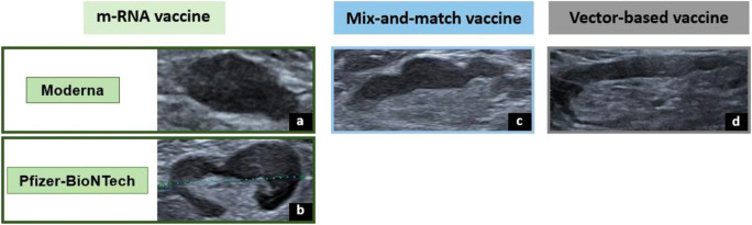
A total of 247 volunteer employees from our center who had received two doses of COVID-19 vaccination were recruited and followed prospectively. Axillary ultrasound of the ipsilateral vaccinated arm was performed the week after receiving the second dose to analyze lymph node features (number, long-axis, cortical thickness, morphology, and vascular imaging). Axillary lymphadenopathy resulting from four vaccination protocols-mRNA (BNT162b2, mRNA-1273), ChAdOx1-S, and mix-and-match-was compared. Analysis was conducted using the Kruskal-Wallis test and post hoc analysis with Bonferroni corrections. Nodal reactogenicity was evaluated for two age groups: young (< 45 years old) and middle-aged ( ≥ 45 years old). All parameters were compared between both groups using an unpaired-sample Student t test. A p value < 0.05 was considered statistically significant.
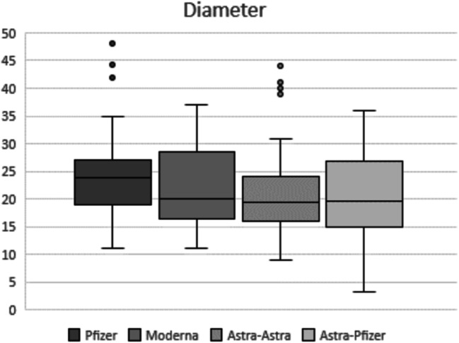
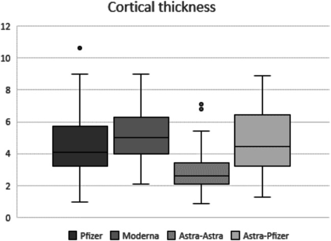
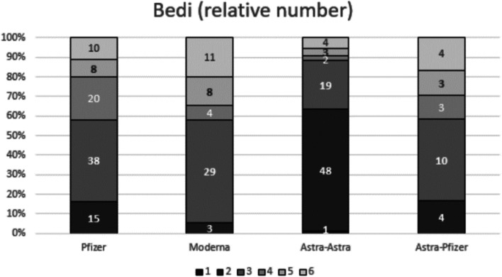
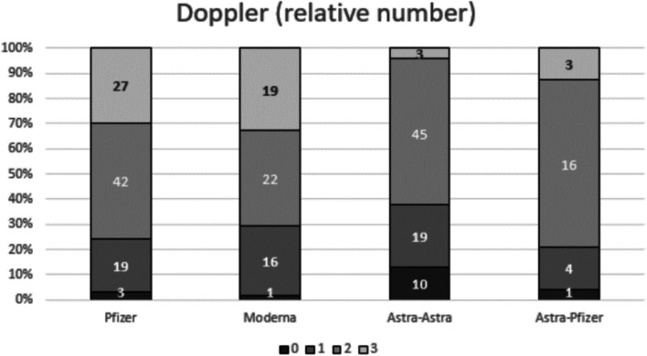
Significantly higher values for total number of visible nodes, cortical thickness, Bedi's classification (p < 0.001), and vascularity (p < 0.05) were observed in mRNA vaccine recipients compared to full ChAdOx1-S protocol recipients. Moreover, mix-and-match protocol recipients showed greater nodal cortical thickness and higher Bedi's classification than full ChAdOx1-S recipients (p < 0.001). Analyses between age groups revealed greater cortical thickness, Bedi's classification, and color Doppler signal in younger patients (p < 0.05).
Nodal parameters of Bedi's classification and cortical thickness were more often increased in mRNA and mix-and-match vaccine recipients when compared to ChAdOx1-S vaccine alone, especially in younger patients.
• Hyperplastic lymphadenopathy was observed more frequently in mRNA and mix-and-match vaccine protocols compared to full vector-based vaccination. • Higher values for cortical thickness, Bedi's classification, and color Doppler signal parameters were identified in younger patients. • Observed lymph node findings normalized in greater than 80% of patients by the third month following vaccination.
评估四种不同 COVID-19 疫苗接种方案两剂后同侧腋窝淋巴结的超声特征,确定这些参数是否随年龄而变化,并描述其在随访影像学上的变化。
我们中心招募了 247 名接受过两剂 COVID-19 疫苗接种的志愿者员工,并进行前瞻性随访。在接受第二剂疫苗后一周,对同侧接种疫苗的手臂进行腋窝超声检查,以分析淋巴结特征(数量、长轴、皮质厚度、形态和血管成像)。比较了四种疫苗接种方案(mRNA [BNT162b2、mRNA-1273]、ChAdOx1-S 和混合接种)引起的腋窝淋巴结病。采用 Kruskal-Wallis 检验和事后 Bonferroni 校正的析因分析进行分析。对两个年龄组(<45 岁和≥45 岁)进行淋巴结反应性评估。使用独立样本 t 检验比较两组间所有参数。p 值<0.05 被认为具有统计学意义。
与完整的 ChAdOx1-S 方案相比,mRNA 疫苗接种者的可见淋巴结总数、皮质厚度、Bedi 分类(p<0.001)和血管生成(p<0.05)显著增加。此外,混合接种方案的淋巴结皮质厚度和 Bedi 分类高于完整 ChAdOx1-S 方案(p<0.001)。两组年龄组间的分析显示,年轻患者的皮质厚度、Bedi 分类和彩色多普勒信号更高(p<0.05)。
与单独使用 ChAdOx1-S 疫苗相比,mRNA 和混合接种疫苗方案的淋巴结参数(Bedi 分类和皮质厚度)更常增加,尤其是在年轻患者中。
•与全载体疫苗相比,mRNA 和混合接种方案更常观察到增生性淋巴结病。•年轻患者的皮质厚度、Bedi 分类和彩色多普勒信号参数值较高。•接种疫苗后 3 个月内,超过 80%的患者的淋巴结发现正常化。