Cocco Giulio, Delli Pizzi Andrea, Fabiani Stefano, Cocco Nino, Boccatonda Andrea, Frisone Alessio, Scarano Antonio, Schiavone Cosima
Unit of Ultrasound in Internal Medicine, Department of Medicine and Science of Aging, "G. d'Annunzio" University, 66100 Chieti, Italy.
Department of Neurosciences, Imaging and Clinical Sciences, "G. d'Annunzio" University, 66100 Chieti, Italy.
Biology (Basel). 2021 Jul 12;10(7):652. doi: 10.3390/biology10070652.
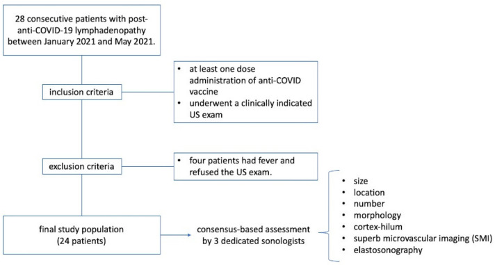
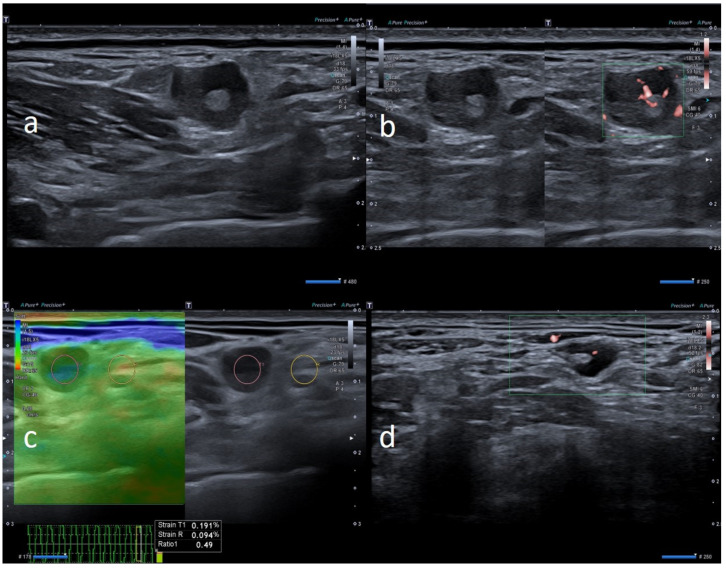
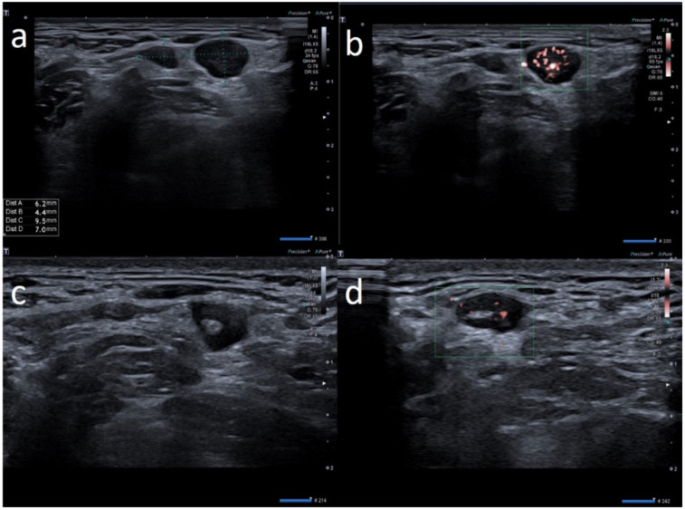
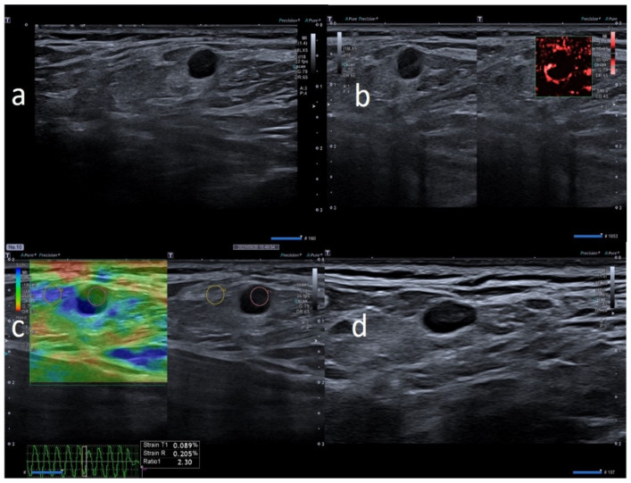
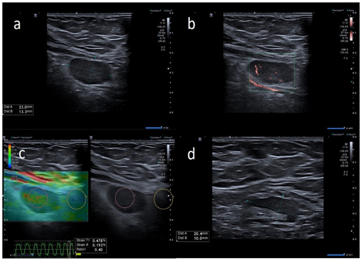
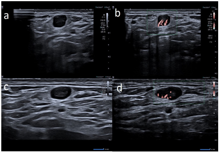
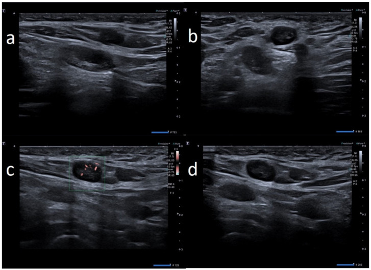
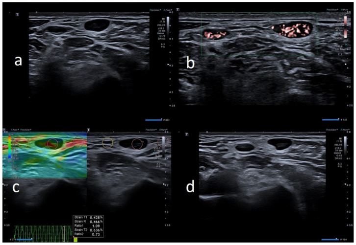
: Post-anti-COVID-19 vaccine lymphadenopathy has recently been described in the literature. In this study, we investigated the multiparametric US findings of patients with post-vaccine lymphadenopathy and compared these findings among different anti-COVID-19 vaccines. : We retrospectively evaluated 24 patients who underwent US between January and May 2021 due to post-anti-COVID-19 lymphadenopathy. The presence, size, location, number, morphology, cortex-hilum, superb microvascular imaging (SMI) and elastosonography of lymph nodes were assessed. Descriptive statistics were calculated and differences among anti-COVID-19 vaccines were analyzed using the Kruskal-Wallis test. A -value ≤ 0.05 was considered statistically significant. : Sixty-six nodes were assessed. They were axillary (mean 1.6 cm ± 0.16) in 11 patients (45.8%) and supraclavicular (mean 0.9 cm ± 0.19) in 13 patients (54.2%). In 20 patients (83.3%), the number of nodes was ≤3. Prevalent US features included oval morphology (18, 75%), asymmetric cortex with hilum evidence (9, 37.5%), central and peripheral vascular signals (12, 50%) at SMI and elastosonography patterns similar to the surrounding tissue (15, 71.4%). No significant differences among the three anti-COVID-19 vaccines were observed ( > 0.05). : Anti-COVID-19 vaccines may present lymphadenopathy with "worrisome" US features regarding size, shape, morphology, cortex-hilum, SMI and elastosonography. An awareness of the patient's history and US findings may help in the early recognition of this clinical scenario and in the appropriate selection of patients for a short-term US follow-up.
: 抗新冠病毒疫苗接种后淋巴结病最近在文献中有所描述。在本研究中,我们调查了接种疫苗后淋巴结病患者的多参数超声检查结果,并比较了不同抗新冠病毒疫苗之间的这些结果。: 我们回顾性评估了2021年1月至5月因抗新冠病毒疫苗接种后淋巴结病接受超声检查的24例患者。评估了淋巴结的存在、大小、位置、数量、形态、皮质-髓质、超微血管成像(SMI)和弹性成像。计算描述性统计量,并使用Kruskal-Wallis检验分析抗新冠病毒疫苗之间的差异。P值≤0.05被认为具有统计学意义。: 共评估了66个淋巴结。11例患者(45.8%)的淋巴结位于腋窝(平均1.6 cm±0.16),13例患者(54.2%)的淋巴结位于锁骨上(平均0.9 cm±0.19)。20例患者(83.3%)的淋巴结数量≤3个。常见的超声特征包括椭圆形形态(占75%,18个)、有髓质证据的不对称皮质(占37.5%,9个)、SMI时的中央和周边血管信号(占50%,12个)以及与周围组织相似的弹性成像模式(占71.4%,15个)。三种抗新冠病毒疫苗之间未观察到显著差异(P>0.05)。: 抗新冠病毒疫苗接种后可能出现具有大小、形状、形态、皮质-髓质、SMI和弹性成像等“令人担忧”超声特征的淋巴结病。了解患者病史和超声检查结果有助于早期识别这种临床情况,并有助于适当选择患者进行短期超声随访。