Abudula Maidinaimu, Fan Xiaodan, Zhang Jing, Li Jiajie, Zhou Xiaoming, Chen Yichen
Medical School, Ningbo University, Ningbo, China.
Department of Gynecology, Ningbo Women and Children's Hospital, Ningbo, China.
Front Cell Dev Biol. 2022 Apr 26;10:824075. doi: 10.3389/fcell.2022.824075. eCollection 2022.
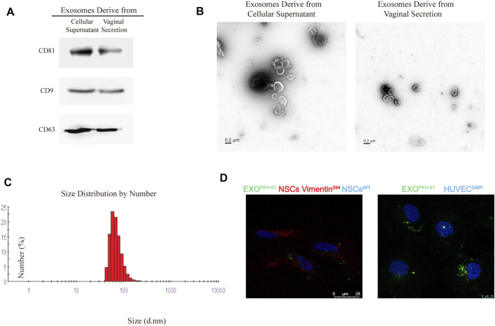
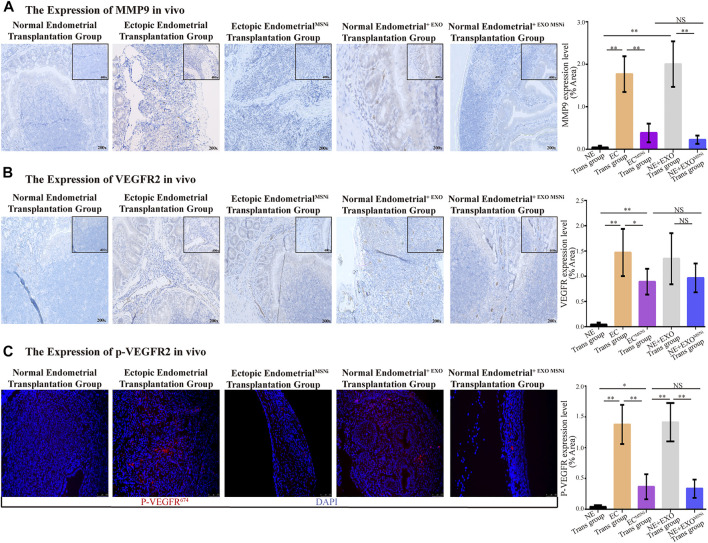
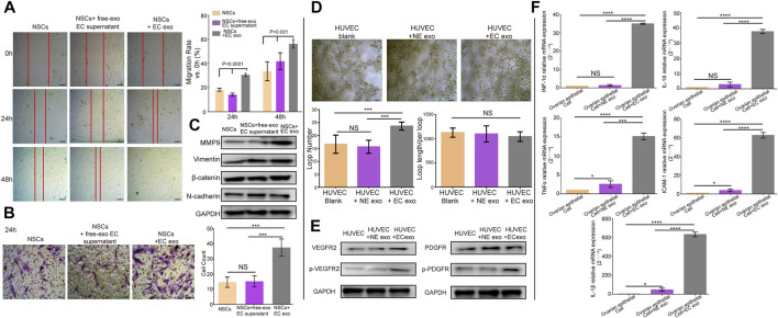
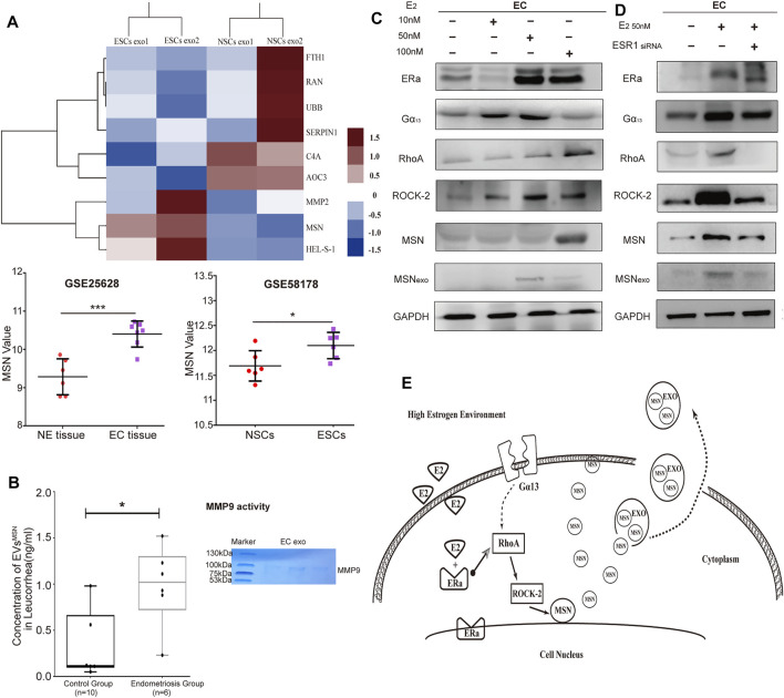
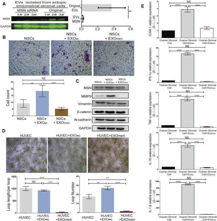
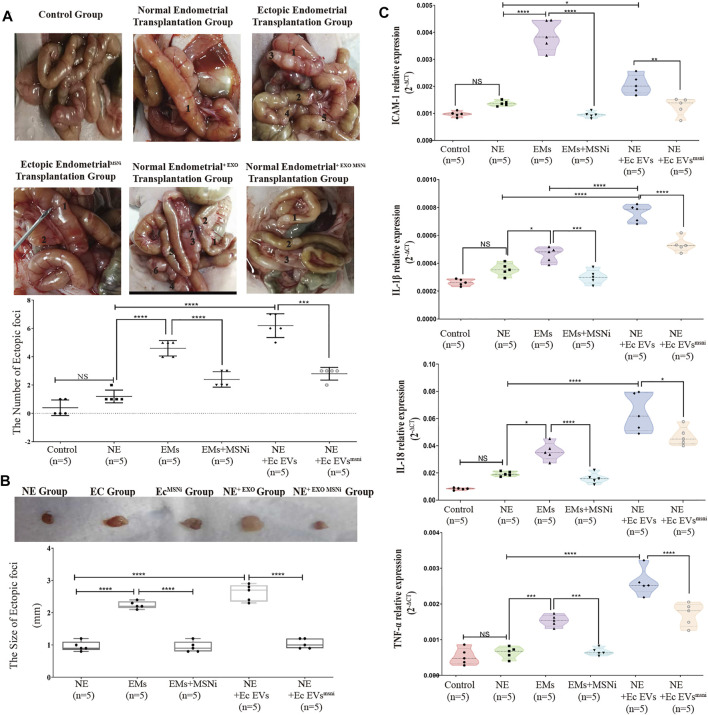
Endometriosis (EMs) is the most common gynaecological disorder with its etiology and/or pathophysiology remains enigmatic. Recent studies showed that extracellular vesicles (EVs), exosomes in particular, play a critical role in developing various clinical disorders. However, the implication of exosomes in endometriosis progression has not been well elucidated. The ectopic stromal cellular exosomes (eEVs) were assessed by transwell assay, scratch tests, tube formation assay, western blot, and qRT-PCR analysis. Protein expression profiles of exosomes in endometrial tissue and vaginal discharge collected from patients with EMS and healthy donors were analysed by Mass spectrometry. siRNA interference technology was used to inhibit the expression of exosomal protein for the functional analysis in . Finally, experiments were performed to validate the results that we observed in EMs mouse model. , we discovered that eEVs improved NSC migratory potential by upregulating MMP9 expression and activity. eEVs also aided angiogenesis and elevated the expression of inflammatory cytokines in ovarian epithelial cells, according to our findings. Moesin (MSN) levels in ESC exosomes were substantially greater than in NSC exosomes (1.22e±5.58e vs. 6.605e±4.574e, LFQ intensity), as shown by protein mass spectrometry and bioinformatics analysis. In ectopic stromal cells, ERa receptors stimulated the RhoA/Rock-2/MSN pathway. We discovered that downregulating exosomal moesin reduced NSC migration (about 3-fold change) and MMP9 expression (about 2-fold change). On the other hand, Exomsni inhibited angiogenesis and inflammatory cytokine release. the result of immunohistochemistry and immunofluorescence demonstrated that exosomal MSN substantially modified the expression of MM9, VEGFR and p-VEGFR in polyclonal lesions. In addition, we discovered an elevation in the expression of proinflammatory factors in the surrounding tissue. Exosomal MSN derived from ectopic stromal cells can contribute to endometriosis progression by mediating the construction of a "migration-vascularization-inflammation" loop in the ectopic environment.
子宫内膜异位症(EMs)是最常见的妇科疾病,其病因和/或病理生理学仍然不明。最近的研究表明,细胞外囊泡(EVs),尤其是外泌体,在各种临床疾病的发展中起关键作用。然而,外泌体在子宫内膜异位症进展中的作用尚未得到充分阐明。通过Transwell实验、划痕试验、管形成试验、蛋白质印迹和qRT-PCR分析评估异位基质细胞外泌体(eEVs)。通过质谱分析从EMS患者和健康供体收集的子宫内膜组织和阴道分泌物中外泌体的蛋白质表达谱。使用siRNA干扰技术抑制外泌体蛋白的表达以进行功能分析。最后,在EMs小鼠模型中进行实验以验证我们观察到的结果。我们发现,eEVs通过上调MMP9的表达和活性来提高神经干细胞(NSC)的迁移潜力。根据我们的研究结果,eEVs还促进血管生成并提高卵巢上皮细胞中炎性细胞因子的表达。蛋白质质谱和生物信息学分析表明,子宫内膜基质细胞(ESC)外泌体中的膜突蛋白(MSN)水平显著高于NSC外泌体(1.22e±5.58e对6.605e±4.574e,LFQ强度)。在异位基质细胞中,雌激素受体α(ERa)激活RhoA/ROCK-2/MSN信号通路。我们发现下调外泌体膜突蛋白可减少NSC迁移(约3倍变化)和MMP9表达(约2倍变化)。另一方面,Exomsni抑制血管生成和炎性细胞因子释放。免疫组织化学和免疫荧光结果表明,外泌体MSN显著改变多克隆病变中MM9、血管内皮生长因子受体(VEGFR)和磷酸化VEGFR(p-VEGFR)的表达。此外,我们发现周围组织中促炎因子的表达升高。异位基质细胞来源的外泌体MSN可通过介导异位环境中“迁移-血管生成-炎症”环路的构建促进子宫内膜异位症的进展。