Kras Katarzyna, Rudyk Halyna, Muszyński Siemowit, Tomaszewska Ewa, Dobrowolski Piotr, Kushnir Volodymyr, Muzyka Viktor, Brezvyn Oksana, Arciszewski Marcin B, Kotsyumbas Ihor
Department of Animal Anatomy and Histology, University of Life Sciences in Lublin, 12 Akademicka St., 20-950 Lublin, Poland.
State Scientific Research Control Institute of Veterinary Medicinal Products and Feed Additives, Donetska St. 11, 79000 Lviv, Ukraine.
Animals (Basel). 2022 Apr 19;12(9):1055. doi: 10.3390/ani12091055.
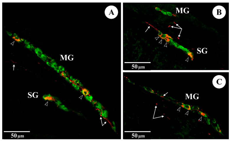
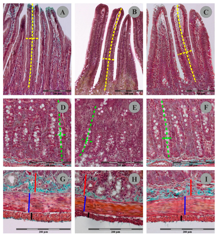
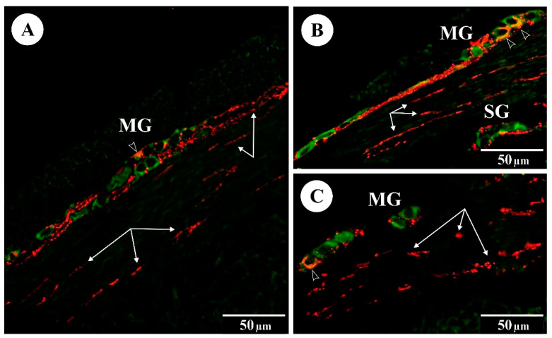
Fumonisins (FBs), including fumonisin B1 and B2 produced by the fungus , are widespread mycotoxins contaminating crop plants as well as processed food. The aim of the experiment was to determine whether the exposure of 5-week-old pregnant rats to FBs at 60 mg/kg b.w. (group FB) or 90 mg/kg b.w. (group FB) results in morphological changes in the duodenum of weaned offspring, particularly the enteric nervous system (ENS). In addition, the levels of expression of galanin and vasoactive intestinal polypeptide (VIP) in the ENS were analysed by immunofluorescence in the control and experimental groups of animals. No significant morphological changes in the thickness of the muscle layer or submucosa of the duodenum were noted in group FB or FB. In group FB (but not FB), there was a significant increase in the width of the villi and in the density of the intestinal crypts. Immunofluorescence analysis using neuronal marker Hu C/D showed no significant changes in group FB or FB in the morphology of the duodenal ENS, i.e., the myenteric plexus (MP) and submucosal plexus (SP), in terms of the density of enteric ganglia in the MP and SP, surface area of MP and SP ganglia, length and width of MP and SP ganglia, surface area of myenteric and submucosal neurons, diameter of myenteric and submucosal neurons, density of myenteric and submucosal neurons, and number of myenteric and submucosal neurons per ganglion. In both groups, there was an increase (relative to the control) in the percentage of Hu C/D-IR/VIP-IR (IR-immunoreactive) and Hu C/D-IR/galanin-IR myenteric and submucosal neurons in the ganglia of both the MP and SP of the duodenum. In addition, in groups FB and FB, there was an increase in the number of nerve fibres showing expression of VIP and galanin in the mucosa, submucosa and circular muscle layer of the duodenum. The results indicate that prenatal exposure to FBs does not significantly alter the histological structure of the duodenum (including the ENS) in the weaned offspring. The changes observed in the chemical code of the myenteric and submucosal neurons in both experimental groups suggest harmful activity of FBs, which may translate into activation of repair mechanisms via overexpression of neuroprotective neuropeptides (VIP and galanin).
伏马毒素(FBs),包括由真菌产生的伏马毒素B1和B2,是广泛存在的霉菌毒素,会污染农作物以及加工食品。本实验的目的是确定5周龄的怀孕大鼠以60毫克/千克体重(FB组)或90毫克/千克体重(FB组)接触伏马毒素是否会导致断奶后代十二指肠出现形态变化,特别是肠神经系统(ENS)。此外,通过免疫荧光分析对照组和实验组动物ENS中甘丙肽和血管活性肠肽(VIP)的表达水平。在FB组或FB组中,未观察到十二指肠肌层或黏膜下层厚度有明显的形态变化。在FB组(而非FB组)中,绒毛宽度和肠隐窝密度显著增加。使用神经元标记物Hu C/D进行的免疫荧光分析显示,在FB组或FB组中,十二指肠ENS,即肌间神经丛(MP)和黏膜下神经丛(SP)的形态,在MP和SP中肠神经节的密度、MP和SP神经节的表面积、MP和SP神经节的长度和宽度、肌间和黏膜下神经元的表面积、肌间和黏膜下神经元的直径、肌间和黏膜下神经元的密度以及每个神经节中肌间和黏膜下神经元的数量方面均无显著变化。在两组中,十二指肠MP和SP神经节中Hu C/D免疫反应阳性(IR)/VIP免疫反应阳性以及Hu C/D免疫反应阳性/甘丙肽免疫反应阳性的肌间和黏膜下神经元的百分比相对于对照组均有所增加。此外,在FB组和FB组中,十二指肠黏膜、黏膜下层和环形肌层中显示VIP和甘丙肽表达的神经纤维数量增加。结果表明,产前接触伏马毒素不会显著改变断奶后代十二指肠(包括ENS)的组织结构。在两个实验组中观察到的肌间和黏膜下神经元化学编码的变化表明伏马毒素具有有害活性,这可能通过神经保护神经肽(VIP和甘丙肽)的过表达转化为修复机制的激活。