• 文献检索
  • 文档翻译
  • 深度研究
  • 学术资讯
  • Suppr Zotero 插件Zotero 插件
  • 邀请有礼
  • 套餐&价格
  • 历史记录
应用&插件
Suppr Zotero 插件Zotero 插件浏览器插件Mac 客户端Windows 客户端微信小程序
定价
高级版会员购买积分包购买API积分包
服务
文献检索文档翻译深度研究API 文档MCP 服务
关于我们
关于 Suppr公司介绍联系我们用户协议隐私条款
关注我们

Suppr 超能文献

核心技术专利:CN118964589B侵权必究
粤ICP备2023148730 号-1Suppr @ 2026

文献检索

告别复杂PubMed语法,用中文像聊天一样搜索,搜遍4000万医学文献。AI智能推荐,让科研检索更轻松。

立即免费搜索

文件翻译

保留排版,准确专业,支持PDF/Word/PPT等文件格式,支持 12+语言互译。

免费翻译文档

深度研究

AI帮你快速写综述,25分钟生成高质量综述,智能提取关键信息,辅助科研写作。

立即免费体验

直肠癌中的淋巴结评估:我们目前的状况与未来展望

Lymph Nodes Evaluation in Rectal Cancer: Where Do We Stand and Future Perspective.

作者信息

Borgheresi Alessandra, De Muzio Federica, Agostini Andrea, Ottaviani Letizia, Bruno Alessandra, Granata Vincenza, Fusco Roberta, Danti Ginevra, Flammia Federica, Grassi Roberta, Grassi Francesca, Bruno Federico, Palumbo Pierpaolo, Barile Antonio, Miele Vittorio, Giovagnoni Andrea

机构信息

Department of Clinical, Special and Dental Sciences, University Politecnica delle Marche, 60121 Ancona, Italy.

Department of Medicine and Health Sciences "V. Tiberio", University of Molise, 86100 Campobasso, Italy.

出版信息

J Clin Med. 2022 May 5;11(9):2599. doi: 10.3390/jcm11092599.

DOI:10.3390/jcm11092599
PMID:35566723
原文链接:https://pmc.ncbi.nlm.nih.gov/articles/PMC9104021/
Abstract

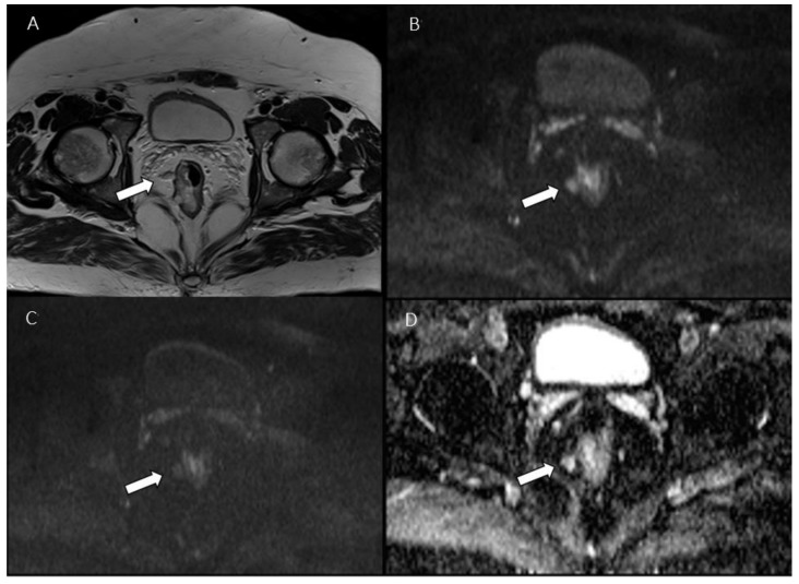
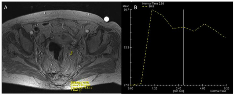
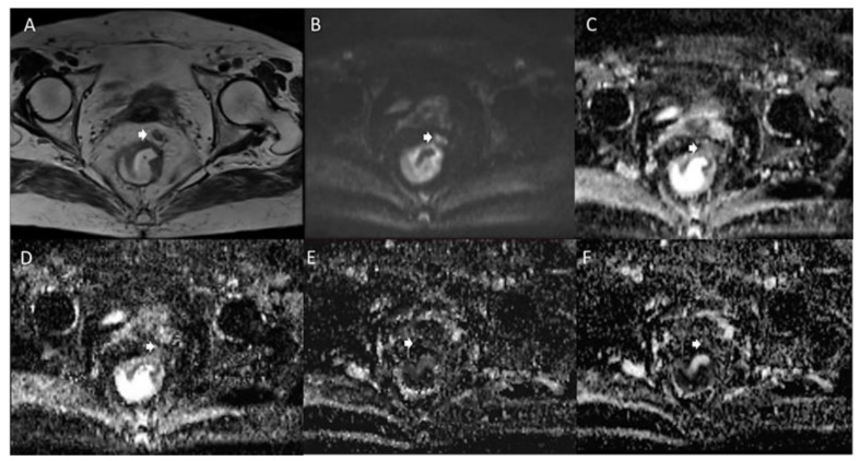
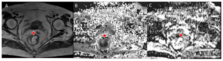
The assessment of nodal involvement in patients with rectal cancer (RC) is fundamental in disease management. Magnetic Resonance Imaging (MRI) is routinely used for local and nodal staging of RC by using morphological criteria. The actual dimensional and morphological criteria for nodal assessment present several limitations in terms of sensitivity and specificity. For these reasons, several different techniques, such as Diffusion Weighted Imaging (DWI), Intravoxel Incoherent Motion (IVIM), Diffusion Kurtosis Imaging (DKI), and Dynamic Contrast Enhancement (DCE) in MRI have been introduced but still not fully validated. Positron Emission Tomography (PET)/CT plays a pivotal role in the assessment of LNs; more recently PET/MRI has been introduced. The advantages and limitations of these imaging modalities will be provided in this narrative review. The second part of the review includes experimental techniques, such as iron-oxide particles (SPIO), and dual-energy CT (DECT). Radiomics analysis is an active field of research, and the evidence about LNs in RC will be discussed. The review also discusses the different recommendations between the European and North American guidelines for the evaluation of LNs in RC, from anatomical considerations to structured reporting.

摘要

评估直肠癌(RC)患者的淋巴结受累情况是疾病管理的基础。磁共振成像(MRI)通常利用形态学标准对RC进行局部和淋巴结分期。淋巴结评估的实际尺寸和形态学标准在敏感性和特异性方面存在若干局限性。基于这些原因,已引入了几种不同的技术,如MRI中的扩散加权成像(DWI)、体素内不相干运动(IVIM)、扩散峰度成像(DKI)和动态对比增强(DCE),但仍未得到充分验证。正电子发射断层扫描(PET)/CT在淋巴结评估中起着关键作用;最近又引入了PET/MRI。本叙述性综述将介绍这些成像方式的优缺点。综述的第二部分包括实验技术,如氧化铁颗粒(SPIO)和双能CT(DECT)。放射组学分析是一个活跃的研究领域,将讨论有关RC中淋巴结的证据。该综述还讨论了欧洲和北美指南在RC淋巴结评估方面的不同建议,从解剖学考虑到结构化报告。

https://cdn.ncbi.nlm.nih.gov/pmc/blobs/d01b/9104021/20181a6be9b2/jcm-11-02599-g007.jpg
https://cdn.ncbi.nlm.nih.gov/pmc/blobs/d01b/9104021/d62937135558/jcm-11-02599-g001.jpg
https://cdn.ncbi.nlm.nih.gov/pmc/blobs/d01b/9104021/73f89f405c2c/jcm-11-02599-g002.jpg
https://cdn.ncbi.nlm.nih.gov/pmc/blobs/d01b/9104021/09b87941512a/jcm-11-02599-g003.jpg
https://cdn.ncbi.nlm.nih.gov/pmc/blobs/d01b/9104021/61f8a00ef38d/jcm-11-02599-g004.jpg
https://cdn.ncbi.nlm.nih.gov/pmc/blobs/d01b/9104021/efa14a4cabce/jcm-11-02599-g005.jpg
https://cdn.ncbi.nlm.nih.gov/pmc/blobs/d01b/9104021/7bc7db7553c9/jcm-11-02599-g006.jpg
https://cdn.ncbi.nlm.nih.gov/pmc/blobs/d01b/9104021/20181a6be9b2/jcm-11-02599-g007.jpg

相似文献

1
Lymph Nodes Evaluation in Rectal Cancer: Where Do We Stand and Future Perspective.直肠癌中的淋巴结评估:我们目前的状况与未来展望
J Clin Med. 2022 May 5;11(9):2599. doi: 10.3390/jcm11092599.
2
Histogram models based on intravoxel incoherent motion diffusion-weighted imaging to predict nodal staging of rectal cancer.基于体素内不相干运动扩散加权成像的直方图模型预测直肠癌淋巴结分期。
Eur J Radiol. 2021 Sep;142:109869. doi: 10.1016/j.ejrad.2021.109869. Epub 2021 Jul 20.
3
Assessment of multifocality and axillary nodal involvement in early-stage breast cancer patients using 18F-FDG PET/CT compared to contrast-enhanced and diffusion-weighted magnetic resonance imaging and sentinel node biopsy.与对比增强磁共振成像、扩散加权磁共振成像及前哨淋巴结活检相比,利用18F-FDG PET/CT评估早期乳腺癌患者的多灶性及腋窝淋巴结受累情况。
Acta Radiol. 2015 Aug;56(8):917-23. doi: 10.1177/0284185114539786. Epub 2014 Jul 10.
4
Diffusion and perfusion MR parameters to assess preoperative short-course radiotherapy response in locally advanced rectal cancer: a comparative explorative study among Standardized Index of Shape by DCE-MRI, intravoxel incoherent motion- and diffusion kurtosis imaging-derived parameters.扩散和灌注磁共振参数评估局部进展期直肠癌术前短程放疗反应:DCE-MRI 标准化形状指数、体素内不相干运动和扩散峰度成像衍生参数的比较探索性研究。
Abdom Radiol (NY). 2019 Nov;44(11):3683-3700. doi: 10.1007/s00261-018-1801-z.
5
Multi-parameter diffusion and perfusion magnetic resonance imaging and radiomics nomogram for preoperative evaluation of aquaporin-1 expression in rectal cancer.多参数扩散和灌注磁共振成像及放射组学列线图在直肠癌水通道蛋白-1 表达术前评估中的应用。
Abdom Radiol (NY). 2022 Apr;47(4):1276-1290. doi: 10.1007/s00261-021-03397-x. Epub 2022 Feb 15.
6
Histogram analysis based on intravoxel incoherent motion diffusion-weighted imaging for determining the perineural invasion status of rectal cancer.基于体素内不相干运动扩散加权成像的直方图分析用于确定直肠癌的神经周围侵犯状态
Quant Imaging Med Surg. 2024 Aug 1;14(8):5358-5372. doi: 10.21037/qims-23-1614. Epub 2024 Jul 26.
7
A Nomogram of Combining IVIM-DWI and MRI Radiomics From the Primary Lesion of Rectal Adenocarcinoma to Assess Nonenlarged Lymph Node Metastasis Preoperatively.基于直肠腺癌原发灶 IVIM-DWI 和 MRI 影像组学构建的列线图模型术前评估未增大淋巴结转移的研究
J Magn Reson Imaging. 2022 Sep;56(3):658-667. doi: 10.1002/jmri.28068. Epub 2022 Jan 28.
8
Preoperative detection of lymphovascular invasion in rectal cancer using intravoxel incoherent motion imaging based on radiomics.基于放射组学的体素内不相干运动成像技术术前检测直肠癌的淋巴管侵犯
Med Phys. 2024 Jan;51(1):179-191. doi: 10.1002/mp.16821. Epub 2023 Nov 6.
9
Intravoxel incoherent motion diffusion-weighted imaging as an adjunct to dynamic contrast-enhanced MRI to improve accuracy of the differential diagnosis of benign and malignant breast lesions.体素内不相干运动扩散加权成像作为动态对比增强磁共振成像的辅助手段,以提高乳腺良恶性病变鉴别诊断的准确性。
Magn Reson Imaging. 2017 Feb;36:175-179. doi: 10.1016/j.mri.2016.10.005. Epub 2016 Oct 11.
10
Higher-order diffusion MRI characterization of mesorectal lymph nodes in rectal cancer.直肠癌中直肠系膜淋巴结的高阶扩散磁共振成像特征
Magn Reson Med. 2020 Jul;84(1):348-364. doi: 10.1002/mrm.28102. Epub 2019 Dec 18.

引用本文的文献

1
Preoperative Assessment of Lymph Node Metastasis in Rectal Cancer Using Deep Learning: Investigating the Utility of Various MRI Sequences.使用深度学习对直肠癌淋巴结转移进行术前评估:探究各种MRI序列的效用
Ann Surg Oncol. 2025 Jun 24. doi: 10.1245/s10434-025-17717-8.
2
Artificial intelligence in magnetic resonance imaging for predicting lymph node metastasis in rectal cancer patients: a meta-analysis.磁共振成像中人工智能用于预测直肠癌患者淋巴结转移的Meta分析
Eur Radiol. 2025 Apr 12. doi: 10.1007/s00330-025-11519-y.
3
Novel deep learning algorithm based MRI radiomics for predicting lymph node metastases in rectal cancer.

本文引用的文献

1
Current controversies in TNM for the radiological staging of rectal cancer and how to deal with them: results of a global online survey and multidisciplinary expert consensus.当前直肠癌放射分期 TNM 中存在的争议及处理方法:全球在线调查和多学科专家共识的结果。
Eur Radiol. 2022 Jul;32(7):4991-5003. doi: 10.1007/s00330-022-08591-z. Epub 2022 Mar 7.
2
Dynamic contrast-enhanced (DCE) imaging: state of the art and applications in whole-body imaging.动态对比增强(DCE)成像:全景成像的现状与应用。
Jpn J Radiol. 2022 Apr;40(4):341-366. doi: 10.1007/s11604-021-01223-4. Epub 2021 Dec 24.
3
Biparametric MRI with simplified PI-RADS (S-PI-RADS) for prostate cancer detection and management: what do radiologist need to know.
基于新型深度学习算法的MRI影像组学用于预测直肠癌淋巴结转移
Sci Rep. 2025 Apr 9;15(1):12089. doi: 10.1038/s41598-025-96618-y.
4
PET/MRI is superior to PET/CT in detecting oesophago and gastric carcinomas: a meta-analysis.PET/MRI在检测食管癌和胃癌方面优于PET/CT:一项荟萃分析。
Cancer Imaging. 2025 Apr 7;25(1):50. doi: 10.1186/s40644-025-00871-3.
5
A pelvis MR transformer-based deep learning model for predicting lung metastases risk in patients with rectal cancer.一种基于骨盆磁共振成像的变压器深度学习模型,用于预测直肠癌患者的肺转移风险。
Front Oncol. 2025 Feb 6;15:1496820. doi: 10.3389/fonc.2025.1496820. eCollection 2025.
6
Metastatic Status and Dissection Effect of Regional/Extraregional Lymph Nodes in Japanese Patients with Squamous Cell Carcinoma of the Anal Canal: A Multicenter Retrospective Cohort Study.日本肛管鳞状细胞癌患者区域/区域外淋巴结的转移状态及清扫效果:一项多中心回顾性队列研究
J Anus Rectum Colon. 2025 Jan 25;9(1):33-40. doi: 10.23922/jarc.2024-039. eCollection 2025.
7
Role of Artificial Intelligence in MRI-Based Rectal Cancer Staging: A Systematic Review.人工智能在基于磁共振成像的直肠癌分期中的作用:一项系统综述
Cureus. 2024 Dec 22;16(12):e76185. doi: 10.7759/cureus.76185. eCollection 2024 Dec.
8
State-of-the-art performance of deep learning methods for pre-operative radiologic staging of colorectal cancer lymph node metastasis: a scoping review.深度学习方法在结直肠癌淋巴结转移术前放射学分期中的前沿表现:一项范围综述
BMJ Open. 2024 Dec 2;14(12):e086896. doi: 10.1136/bmjopen-2024-086896.
9
MRI for Rectal Cancer: Updates and Controversies- Expert Panel Narrative Review.直肠癌的磁共振成像:最新进展与争议——专家小组叙述性综述
AJR Am J Roentgenol. 2025 Jun;224(6):e2431523. doi: 10.2214/AJR.24.31523. Epub 2024 Sep 25.
10
The Role of Lateral Pelvic Lymph Node Dissection in Middle and Lower Rectal Cancer (Stage II or III): A Literature Review.侧方盆腔淋巴结清扫术在中低位直肠癌(II期或III期)中的作用:文献综述
Cureus. 2024 Aug 22;16(8):e67526. doi: 10.7759/cureus.67526. eCollection 2024 Aug.
用于前列腺癌检测与管理的简化前列腺影像报告和数据系统(S-PI-RADS)双参数磁共振成像:放射科医生需要了解什么。
Radiol Med. 2021 Dec;126(12):1660-1661. doi: 10.1007/s11547-021-01343-x. Epub 2021 Dec 17.
4
Delta radiomics: a systematic review.德尔塔放射组学:系统评价。
Radiol Med. 2021 Dec;126(12):1571-1583. doi: 10.1007/s11547-021-01436-7. Epub 2021 Dec 4.
5
Intrahepatic cholangiocarcinoma and its differential diagnosis at MRI: how radiologist should assess MR features.MRI 下肝内胆管细胞癌及其鉴别诊断:放射科医生应如何评估 MRI 特征。
Radiol Med. 2021 Dec;126(12):1584-1600. doi: 10.1007/s11547-021-01428-7. Epub 2021 Nov 29.
6
Diagnostic accuracy of dynamic contrast-enhanced magnetic resonance imaging for distinguishing pseudoprogression from glioma recurrence: a meta-analysis.动态对比增强磁共振成像鉴别假性进展与脑胶质瘤复发的诊断准确性:一项荟萃分析。
Chin Med J (Engl). 2021 Oct 14;134(21):2535-2543. doi: 10.1097/CM9.0000000000001445.
7
Structured reporting of computed tomography in the staging of colon cancer: a Delphi consensus proposal.结直肠癌分期 CT 检查的结构化报告:德尔菲共识提案。
Radiol Med. 2022 Jan;127(1):21-29. doi: 10.1007/s11547-021-01418-9. Epub 2021 Nov 6.
8
Radiomics and Digital Image Texture Analysis in Oncology (Review).肿瘤放射组学和数字图像纹理分析(综述)。
Sovrem Tekhnologii Med. 2021;13(2):97-104. doi: 10.17691/stm2021.13.2.11. Epub 2021 Jan 1.
9
Gastrointestinal neuroendocrine neoplasms (GI-NENs): hot topics in morphological, functional, and prognostic imaging.胃肠道神经内分泌肿瘤(GI-NENs):形态学、功能学和预后评估影像学的热点话题。
Radiol Med. 2021 Dec;126(12):1497-1507. doi: 10.1007/s11547-021-01408-x. Epub 2021 Aug 24.
10
Computed tomography-based radiomics approach in pancreatic tumors characterization.基于计算机断层扫描的放射组学方法在胰腺肿瘤特征分析中的应用
Radiol Med. 2021 Aug 12. doi: 10.1007/s11547-021-01405-0.