Infection Program and Department of Microbiology, Biomedicine Discovery Institute, Monash University, Melbourne, VIC, 3800, Australia.
Development and Stem Cells Program and Department of Anatomy & Developmental Biology, Biomedicine Discovery Institute, Monash University, Melbourne, VIC, 3800, Australia.
Cell Mol Life Sci. 2022 May 15;79(6):296. doi: 10.1007/s00018-022-04316-z.
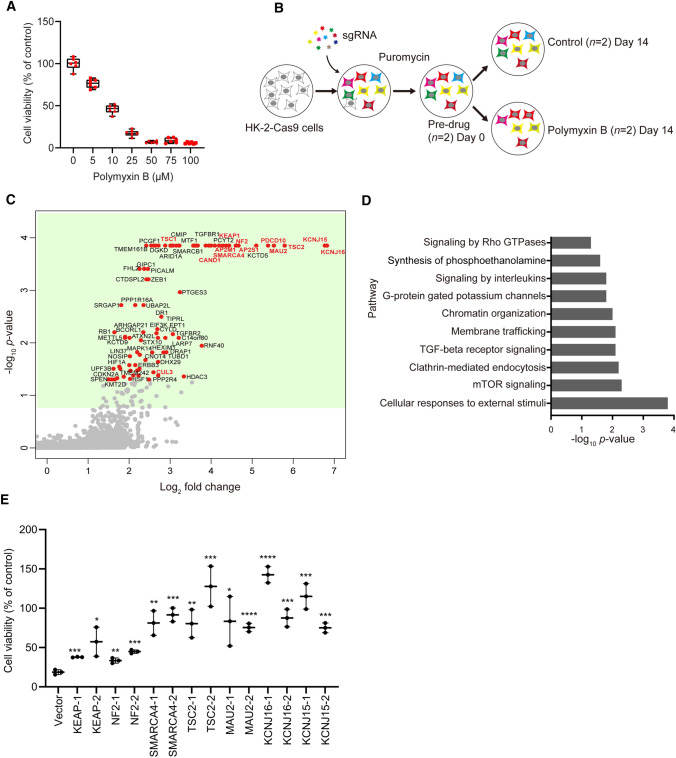
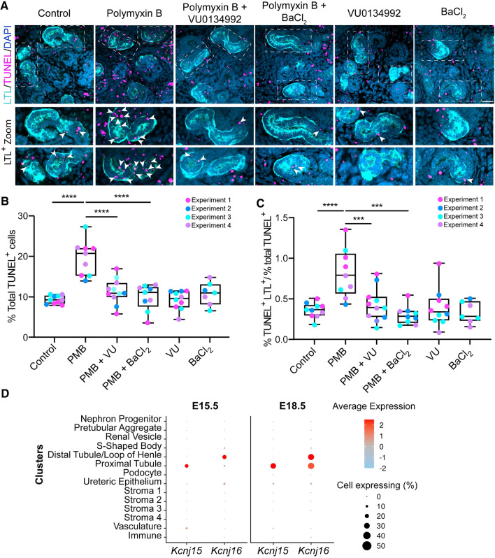
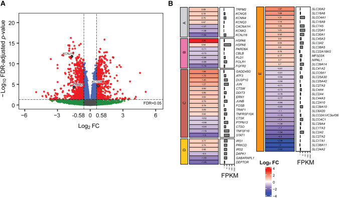
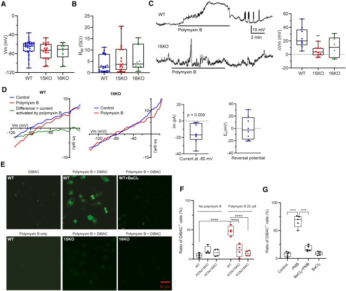
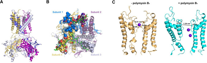
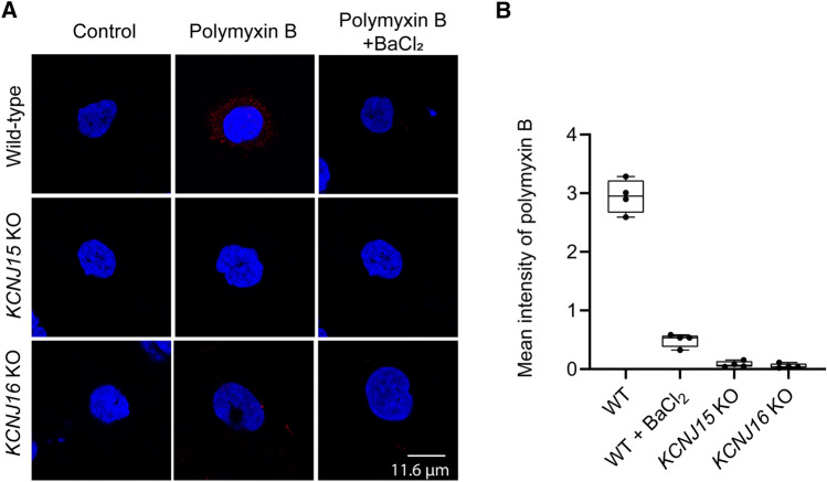
Polymyxin antibiotics are often used as a last-line defense to treat life-threatening Gram-negative pathogens. However, polymyxin-induced kidney toxicity is a dose-limiting factor of paramount importance and can lead to suboptimal treatment. To elucidate the mechanism and develop effective strategies to overcome polymyxin toxicity, we employed a whole-genome CRISPR screen in human kidney tubular HK-2 cells and identified 86 significant genes that upon knock-out rescued polymyxin-induced toxicity. Specifically, we discovered that knockout of the inwardly rectifying potassium channels Kir4.2 and Kir5.1 (encoded by KCNJ15 and KCNJ16, respectively) rescued polymyxin-induced toxicity in HK-2 cells. Furthermore, we found that polymyxins induced cell depolarization via Kir4.2 and Kir5.1 and a significant cellular uptake of polymyxins was evident. All-atom molecular dynamics simulations revealed that polymyxin B spontaneously bound to Kir4.2, thereby increasing opening of the channel, resulting in a potassium influx, and changes of the membrane potential. Consistent with these findings, small molecule inhibitors (BaCl and VU0134992) of Kir potassium channels reduced polymyxin-induced toxicity in cell culture and mouse explant kidney tissue. Our findings provide critical mechanistic information that will help attenuate polymyxin-induced nephrotoxicity in patients and facilitate the design of novel, safer polymyxins.
多黏菌素类抗生素常被用作治疗危及生命的革兰氏阴性病原体的最后防线。然而,多黏菌素诱导的肾毒性是一个至关重要的剂量限制因素,可能导致治疗效果不佳。为了阐明机制并开发克服多黏菌素毒性的有效策略,我们在人肾小管 HK-2 细胞中进行了全基因组 CRISPR 筛选,鉴定出 86 个重要基因,敲除这些基因可挽救多黏菌素诱导的毒性。具体来说,我们发现敲除内向整流钾通道 Kir4.2 和 Kir5.1(分别由 KCNJ15 和 KCNJ16 编码)可挽救 HK-2 细胞中的多黏菌素诱导的毒性。此外,我们发现多黏菌素通过 Kir4.2 和 Kir5.1 诱导细胞去极化,并且多黏菌素的明显细胞摄取是显而易见的。全原子分子动力学模拟表明,多黏菌素 B 可自发结合 Kir4.2,从而增加通道的开放,导致钾内流和膜电位的变化。与这些发现一致,Kir 钾通道的小分子抑制剂(BaCl 和 VU0134992)可减少细胞培养和小鼠肾组织中的多黏菌素诱导的毒性。我们的研究结果提供了关键的机制信息,有助于减轻患者的多黏菌素诱导的肾毒性,并促进新型、更安全的多黏菌素的设计。