University of Toronto, Toronto, Canada.
University of Alberta, Alberta, Canada.
Oncologist. 2022 Sep 2;27(9):e748-e754. doi: 10.1093/oncolo/oyac092.
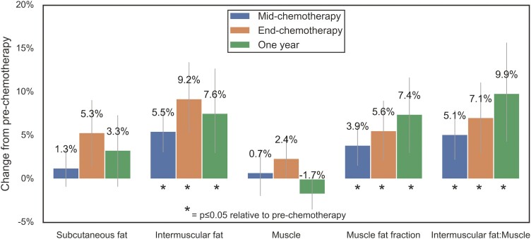
While cardiotoxic chemotherapy is known to negatively impact cardiac function and hemoglobin levels, the impact on skeletal muscle has been understudied among patients. The purpose was to longitudinally characterize myosteatosis (muscle fat), skeletal muscle metabolism, and oxygen (O2) consumption during cardiotoxic chemotherapy for breast cancer.
Thirty-four patients with stage I-III breast cancer were enrolled before trastuzumab-containing and/or anthracycline-containing chemotherapy. We used magnetic resonance imaging to non-invasively quantify thigh myosteatosis (fat-water imaging), and lower leg metabolism (31P spectroscopy), O2 consumption (custom techniques), and peak power output during single-leg plantarflexion exercise at pre-, mid-, end-chemotherapy, and 1-year. We also measured pulmonary VO2peak and maximal leg press strength.
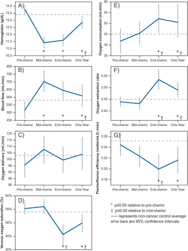
During chemotherapy, VO2peak and leg press strength decreased while peak plantarflexion power output was maintained. At mid-chemotherapy, hemoglobin decreased (16%) and lower leg blood flow increased (37%) to maintain lower leg O2 delivery; exercise Pi:PCr and myosteatosis increased. Between mid- and end-chemotherapy, lower leg O2 extraction (28%) and O2 consumption (21%) increased, while plantarflexion exercise efficiency (watts/O2 consumed) decreased. At one year, VO2peak and leg press strength returned to pre-chemotherapy levels, but lower leg exercise O2 extraction, consumption and Pi:PCr, and myosteatosis remained elevated.
Lower leg skeletal muscle blood flow and O2 extraction adapt to compensate for chemotherapy-related hemoglobin reduction for small muscle mass exercise but are insufficient to maintain large muscle mass exercise (pulmonary VO2peak, leg press strength). The excess O2 required to perform work, increased Pi:PCr ratio and myosteatosis together suggest suppressed fat oxidation during chemotherapy.
已知心脏毒性化疗会对心脏功能和血红蛋白水平产生负面影响,但在接受心脏毒性化疗的患者中,骨骼肌的影响尚未得到充分研究。本研究旨在对乳腺癌患者接受心脏毒性化疗过程中的肌内脂肪(肌肉脂肪)、骨骼肌代谢和耗氧量进行纵向特征描述。
招募了 34 名 I-III 期乳腺癌患者,在接受曲妥珠单抗和/或蒽环类药物化疗前进行检查。我们使用磁共振成像技术无创性地定量大腿肌内脂肪(水-脂肪成像)和小腿代谢(31P 光谱)、耗氧量(定制技术)以及单腿跖屈运动时的峰值功率输出,在化疗前、化疗中、化疗结束和 1 年后进行检测。我们还测量了肺 VO2peak 和最大腿举力量。
在化疗期间,VO2peak 和腿举力量下降,而峰值跖屈功率输出保持不变。在化疗中期,血红蛋白下降(16%),小腿血流量增加(37%),以维持小腿 O2 输送;运动时 Pi:PCr 和肌内脂肪增加。在化疗中期和结束时,小腿 O2 摄取(28%)和 O2 消耗(21%)增加,而跖屈运动效率(消耗的瓦特/O2)下降。在 1 年时,VO2peak 和腿举力量恢复到化疗前水平,但小腿运动时的 O2 摄取、消耗和 Pi:PCr 以及肌内脂肪仍然升高。
小腿骨骼肌血流量和 O2 摄取适应以代偿化疗相关的血红蛋白减少,以维持小肌肉量运动(肺 VO2peak、腿举力量),但不足以维持大肌肉量运动。完成工作所需的额外 O2、增加的 Pi:PCr 比值和肌内脂肪提示,在化疗期间脂肪氧化受到抑制。