• 文献检索
  • 文档翻译
  • 深度研究
  • 学术资讯
  • Suppr Zotero 插件Zotero 插件
  • 邀请有礼
  • 套餐&价格
  • 历史记录
应用&插件
Suppr Zotero 插件Zotero 插件浏览器插件Mac 客户端Windows 客户端微信小程序
定价
高级版会员购买积分包购买API积分包
服务
文献检索文档翻译深度研究API 文档MCP 服务
关于我们
关于 Suppr公司介绍联系我们用户协议隐私条款
关注我们

Suppr 超能文献

核心技术专利:CN118964589B侵权必究
粤ICP备2023148730 号-1Suppr @ 2026

文献检索

告别复杂PubMed语法,用中文像聊天一样搜索,搜遍4000万医学文献。AI智能推荐,让科研检索更轻松。

立即免费搜索

文件翻译

保留排版,准确专业,支持PDF/Word/PPT等文件格式,支持 12+语言互译。

免费翻译文档

深度研究

AI帮你快速写综述,25分钟生成高质量综述,智能提取关键信息,辅助科研写作。

立即免费体验

水合作用介导的 G 蛋白偶联受体激活。

Hydration-mediated G-protein-coupled receptor activation.

机构信息

Department of Chemistry and Biochemistry, University of Arizona, Tucson, AZ 85721.

Laboratory of Biomolecular NMR, St. Petersburg State University, St. Petersburg 199034, Russia.

出版信息

Proc Natl Acad Sci U S A. 2022 May 24;119(21):e2117349119. doi: 10.1073/pnas.2117349119. Epub 2022 May 18.

DOI:10.1073/pnas.2117349119
PMID:35584119
原文链接:https://pmc.ncbi.nlm.nih.gov/articles/PMC9173805/
Abstract

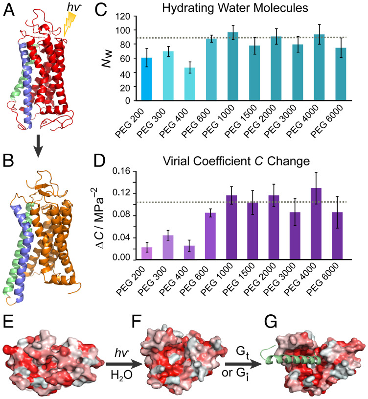
The Rhodopsin family of G-protein–coupled receptors (GPCRs) comprises the targets of nearly a third of all pharmaceuticals. Despite structural water present in GPCR X-ray structures, the physiological relevance of these solvent molecules to rhodopsin signaling remains unknown. Here, we show experimental results consistent with the idea that rhodopsin activation in lipid membranes is coupled to bulk water movements into the protein. To quantify hydration changes, we measured reversible shifting of the metarhodopsin equilibrium due to osmotic stress using an extensive series of polyethylene glycol (PEG) osmolytes. We discovered clear evidence that light activation entails a large influx of bulk water (∼80–100 molecules) into the protein, giving insight into GPCR activation mechanisms. Various size polymer osmolytes directly control rhodopsin activation, in which large solutes are excluded from rhodopsin and dehydrate the protein, favoring the inactive state. In contrast, small osmolytes initially forward shift the activation equilibrium until a quantifiable saturation point is reached, similar to gain-of-function protein mutations. For the limit of increasing osmolyte size, a universal response of rhodopsin to osmotic stress is observed, suggesting it adopts a dynamic, hydrated sponge-like state upon photoactivation. Our results demand a rethinking of the role of water dynamics in modulating various intermediates in the GPCR energy landscape. We propose that besides bound water, an influx of bulk water plays a necessary role in establishing the active GPCR conformation that mediates signaling.

摘要

G 蛋白偶联受体 (GPCR) 中的视紫红质家族包含了近三分之一的药物靶点。尽管 GPCR X 射线结构中存在结构水,但这些溶剂分子对视紫红质信号转导的生理相关性尚不清楚。在这里,我们展示了与以下观点一致的实验结果,即在脂质膜中,视紫红质的激活与大量水分子进入蛋白质有关。为了定量测量水合变化,我们使用一系列广泛的聚乙二醇 (PEG) 渗透剂来测量渗透压应激下视黄醛平衡的可逆移动。我们发现了明确的证据表明,光激活会导致大量的 bulk water(~80-100 个分子)进入蛋白质,从而深入了解 GPCR 激活机制。各种大小的聚合物渗透剂直接控制视紫红质的激活,其中大溶质被排除在视紫红质之外并使蛋白质脱水,有利于非活性状态。相比之下,小渗透剂最初会向前移动激活平衡,直到达到可量化的饱和点,类似于获得功能的蛋白质突变。对于渗透剂尺寸不断增加的极限,视紫红质对渗透压应激的反应是一致的,这表明它在光激活后会采取一种动态的、水合的海绵状状态。我们的结果要求重新思考水动力学在调节 GPCR 能量景观中的各种中间物的作用。我们提出,除了结合水之外,大量水的流入在建立介导信号转导的活性 GPCR 构象中起着必要的作用。

https://cdn.ncbi.nlm.nih.gov/pmc/blobs/23ef/9173805/c7f6c6bda4d4/pnas.2117349119fig04.jpg
https://cdn.ncbi.nlm.nih.gov/pmc/blobs/23ef/9173805/ae82ac3e2109/pnas.2117349119fig01.jpg
https://cdn.ncbi.nlm.nih.gov/pmc/blobs/23ef/9173805/30284b10a4b3/pnas.2117349119fig02.jpg
https://cdn.ncbi.nlm.nih.gov/pmc/blobs/23ef/9173805/80f26bbcc9fb/pnas.2117349119fig03.jpg
https://cdn.ncbi.nlm.nih.gov/pmc/blobs/23ef/9173805/c7f6c6bda4d4/pnas.2117349119fig04.jpg

相似文献

1
Hydration-mediated G-protein-coupled receptor activation.水合作用介导的 G 蛋白偶联受体激活。
Proc Natl Acad Sci U S A. 2022 May 24;119(21):e2117349119. doi: 10.1073/pnas.2117349119. Epub 2022 May 18.
2
Osmotic stress studies of G-protein-coupled receptor rhodopsin activation.G蛋白偶联受体视紫红质激活的渗透应激研究。
Biophys Chem. 2024 Jan;304:107112. doi: 10.1016/j.bpc.2023.107112. Epub 2023 Sep 26.
3
Activation of the G-Protein-Coupled Receptor Rhodopsin by Water.水对 G 蛋白偶联受体视紫红质的激活作用。
Angew Chem Int Ed Engl. 2021 Feb 1;60(5):2288-2295. doi: 10.1002/anie.202003342. Epub 2020 Nov 27.
4
Agonist-induced conformational changes in bovine rhodopsin: insight into activation of G-protein-coupled receptors.激动剂诱导的牛视紫红质构象变化:深入了解G蛋白偶联受体的激活
J Mol Biol. 2008 Oct 3;382(2):539-55. doi: 10.1016/j.jmb.2008.06.084. Epub 2008 Jul 7.
5
Relevance of rhodopsin studies for GPCR activation.视紫红质研究对G蛋白偶联受体激活的相关性。
Biochim Biophys Acta. 2014 May;1837(5):674-82. doi: 10.1016/j.bbabio.2013.09.002. Epub 2013 Sep 13.
6
Internal hydration increases during activation of the G-protein-coupled receptor rhodopsin.在G蛋白偶联受体视紫红质激活过程中,内部水合作用增强。
J Mol Biol. 2008 Aug 29;381(2):478-86. doi: 10.1016/j.jmb.2008.05.036. Epub 2008 May 22.
7
Functional water molecules in rhodopsin activation.视紫红质激活中的功能性水分子
J Phys Chem B. 2014 Sep 18;118(37):10863-73. doi: 10.1021/jp505180t. Epub 2014 Sep 8.
8
Equilibrium between metarhodopsin-I and metarhodopsin-II is dependent on the conformation of the third cytoplasmic loop.视紫红质-I与视紫红质-II之间的平衡取决于第三个细胞质环的构象。
J Biol Chem. 2006 Mar 10;281(10):6813-25. doi: 10.1074/jbc.M510175200. Epub 2005 Dec 29.
9
Water permeation through the internal water pathway in activated GPCR rhodopsin.水通过活化的G蛋白偶联受体视紫红质中的内部水通道渗透。
PLoS One. 2017 May 11;12(5):e0176876. doi: 10.1371/journal.pone.0176876. eCollection 2017.
10
Structure and activation of rhodopsin.视紫红质的结构与激活
Acta Pharmacol Sin. 2012 Mar;33(3):291-9. doi: 10.1038/aps.2011.171. Epub 2012 Jan 23.

引用本文的文献

1
H and Confined Water in Gating in Many Voltage-Gated Potassium Channels: Ion/Water/Counterion/Protein Networks and Protons Added to Gate the Channel.许多电压门控钾通道门控中的H与受限水:离子/水/抗衡离子/蛋白质网络以及用于门控通道的质子添加
Int J Mol Sci. 2025 Jul 29;26(15):7325. doi: 10.3390/ijms26157325.
2
Histamine-modulated wettability switching in G-protein-coupled receptor inspired nanochannel for potential drug screening and biosensing.用于潜在药物筛选和生物传感的G蛋白偶联受体启发的纳米通道中的组胺调节润湿性切换
Nat Commun. 2025 Feb 20;16(1):1820. doi: 10.1038/s41467-025-57064-6.
3
Detection of Putative Ligand Dissociation Pathways in Proteins Using Site-Identification by Ligand Competitive Saturation.

本文引用的文献

1
Hydration Dynamics in Biological Membranes: Emerging Applications of Terahertz Spectroscopy.生物膜中的水动态:太赫兹光谱的新兴应用。
J Phys Chem Lett. 2021 Oct 7;12(39):9697-9709. doi: 10.1021/acs.jpclett.1c02576. Epub 2021 Sep 30.
2
Structures of human mGlu2 and mGlu7 homo- and heterodimers.人类代谢型谷氨酸受体2(mGlu2)和代谢型谷氨酸受体7(mGlu7)同二聚体及异二聚体的结构。
Nature. 2021 Jun;594(7864):589-593. doi: 10.1038/s41586-021-03641-w. Epub 2021 Jun 16.
3
Protein-complex stability in cells and in vitro under crowded conditions.
利用配体竞争饱和法进行位点鉴定来检测蛋白质中假定的配体解离途径
J Chem Inf Model. 2025 Mar 24;65(6):3022-3034. doi: 10.1021/acs.jcim.4c01814. Epub 2024 Dec 27.
4
Hidden water's influence on rhodopsin activation.隐匿水对视紫红质激活的影响。
Biophys J. 2024 Dec 17;123(24):4167-4179. doi: 10.1016/j.bpj.2024.11.012. Epub 2024 Nov 16.
5
Cell swelling enhances ligand-driven β-adrenergic signaling.细胞肿胀增强配体驱动的β-肾上腺素能信号。
Nat Commun. 2024 Sep 7;15(1):7822. doi: 10.1038/s41467-024-52191-y.
6
Influence of the Water Model on the Structure and Interactions of the GPR40 Protein with the Lipid Membrane and the Solvent: Rigid versus Flexible Water Models.水模型对 GPR40 蛋白与脂质膜和溶剂的结构和相互作用的影响:刚性水模型与柔性水模型。
J Chem Theory Comput. 2024 Jul 23;20(14):6369-6387. doi: 10.1021/acs.jctc.4c00571. Epub 2024 Jul 11.
7
Membrane Mimetic-Dependence of GPCR Energy Landscapes.G蛋白偶联受体能量景观的膜模拟依赖性。
bioRxiv. 2023 Oct 19:2023.10.16.562552. doi: 10.1101/2023.10.16.562552.
8
Protons taken hostage: Dynamic H-bond networks of the pH-sensing GPR68.被“挟持”的质子:pH 传感受体 GPR68 的动态氢键网络
Comput Struct Biotechnol J. 2023 Sep 2;21:4370-4384. doi: 10.1016/j.csbj.2023.08.034. eCollection 2023.
9
Three-Dimensional Structural Insights Have Revealed the Distinct Binding Interactions of Agonists, Partial Agonists, and Antagonists with the µ Opioid Receptor.三维结构见解揭示了激动剂、部分激动剂和拮抗剂与μ阿片受体的独特结合相互作用。
Int J Mol Sci. 2023 Apr 11;24(8):7042. doi: 10.3390/ijms24087042.
10
Mechanistic basis of GPCR activation explored by ensemble refinement of crystallographic structures.通过晶体结构的总体精修探索 G 蛋白偶联受体激活的机制基础。
Protein Sci. 2022 Nov;31(11):e4456. doi: 10.1002/pro.4456.
细胞内及拥挤环境下体外的蛋白质复合物稳定性。
Curr Opin Struct Biol. 2021 Feb;66:183-192. doi: 10.1016/j.sbi.2020.10.024. Epub 2020 Dec 4.
4
Membrane Curvature Revisited-the Archetype of Rhodopsin Studied by Time-Resolved Electronic Spectroscopy.膜曲率再探讨——视紫红质原型的时间分辨电子能谱研究
Biophys J. 2021 Feb 2;120(3):440-452. doi: 10.1016/j.bpj.2020.11.007. Epub 2020 Nov 18.
5
Activation of the G-Protein-Coupled Receptor Rhodopsin by Water.水对 G 蛋白偶联受体视紫红质的激活作用。
Angew Chem Int Ed Engl. 2021 Feb 1;60(5):2288-2295. doi: 10.1002/anie.202003342. Epub 2020 Nov 27.
6
Depletion interactions modulate the binding between disordered proteins in crowded environments.耗竭相互作用调节拥挤环境中无规则蛋白质之间的结合。
Proc Natl Acad Sci U S A. 2020 Jun 16;117(24):13480-13489. doi: 10.1073/pnas.1921617117. Epub 2020 Jun 2.
7
Flexible lipid nanomaterials studied by NMR spectroscopy.通过 NMR 光谱研究灵活的脂质纳米材料。
Phys Chem Chem Phys. 2019 Aug 28;21(34):18422-18457. doi: 10.1039/c8cp06179c.
8
Structures of the Rhodopsin-Transducin Complex: Insights into G-Protein Activation.视紫红质-转导蛋白复合物的结构:对 G 蛋白激活的深入了解。
Mol Cell. 2019 Aug 22;75(4):781-790.e3. doi: 10.1016/j.molcel.2019.06.007. Epub 2019 Jul 9.
9
Stochasticity of Biological Soft Matter: Emerging Concepts in Intrinsically Disordered Proteins and Biological Phase Separation.生物软物质的随机性:无序蛋白质和生物相分离中的新兴概念。
Trends Biochem Sci. 2019 Aug;44(8):716-728. doi: 10.1016/j.tibs.2019.03.005. Epub 2019 Apr 22.
10
Diverse GPCRs exhibit conserved water networks for stabilization and activation.多种 G 蛋白偶联受体(GPCRs)表现出保守的水网络以稳定和激活。
Proc Natl Acad Sci U S A. 2019 Feb 19;116(8):3288-3293. doi: 10.1073/pnas.1809251116. Epub 2019 Feb 6.