• 文献检索
  • 文档翻译
  • 深度研究
  • 学术资讯
  • Suppr Zotero 插件Zotero 插件
  • 邀请有礼
  • 套餐&价格
  • 历史记录
应用&插件
Suppr Zotero 插件Zotero 插件浏览器插件Mac 客户端Windows 客户端微信小程序
定价
高级版会员购买积分包购买API积分包
服务
文献检索文档翻译深度研究API 文档MCP 服务
关于我们
关于 Suppr公司介绍联系我们用户协议隐私条款
关注我们

Suppr 超能文献

核心技术专利:CN118964589B侵权必究
粤ICP备2023148730 号-1Suppr @ 2026

文献检索

告别复杂PubMed语法,用中文像聊天一样搜索,搜遍4000万医学文献。AI智能推荐,让科研检索更轻松。

立即免费搜索

文件翻译

保留排版,准确专业,支持PDF/Word/PPT等文件格式,支持 12+语言互译。

免费翻译文档

深度研究

AI帮你快速写综述,25分钟生成高质量综述,智能提取关键信息,辅助科研写作。

立即免费体验

DECOMP 报告:腹部壁成像检查中外科医生期望的答案。

DECOMP Report: Answers surgeons expect from an abdominal wall imaging exam.

机构信息

- Universidade Positivo, Clínica Cirúrgica e Cirurgia Minimamente Invasiva - Curitiba - PR - Brasil.

- Hospital Municipal Lourenço Jorge, Clínica Cirúrgica - Rio de Janeiro - RJ - Brasil.

出版信息

Rev Col Bras Cir. 2022 Apr 27;49:e20223172. doi: 10.1590/0100-6991e-20223172en. eCollection 2022.

DOI:10.1590/0100-6991e-20223172en
PMID:35588534
原文链接:https://pmc.ncbi.nlm.nih.gov/articles/PMC10578831/
Abstract

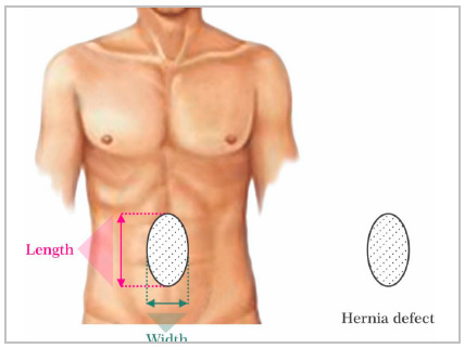
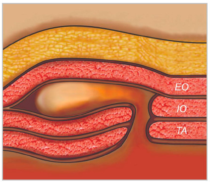
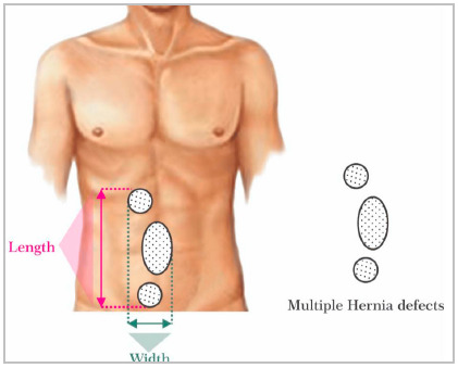
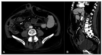
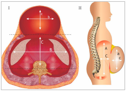
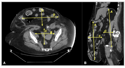
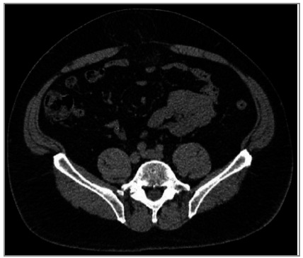
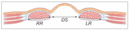
Abdominal wall (AW) hernias are a common problem faced by general surgeons. With an essentially clinical diagnosis, abdominal hernias have been considered a simple problem to be repaired. However, long-term follow-up of patients has shown disappointing results, both in terms of complications and recurrence. In this context, preoperative planning with control of comorbidities and full knowledge of the hernia and its anatomical relationships with the AW has gained increasing attention. Computed tomography (CT) appears to be the best option to determine the precise size and location of abdominal hernias, presence of rectus diastase and/or associated muscle atrophy, as well as the proportion of the hernia in relation to the AW itself. This information might help the surgeon to choose the best surgical technique (open vs MIS), positioning and fixation of the meshes, and eventual need for application of botulinum toxin, preoperative pneumoperitoneum or component separation techniques. Despite the relevance of the findings, they are rarely described in CT scans as radiologists are not used to report findings of the AW as well as to know what information is really needed. For these reasons, we gathered a group of surgeons and radiologists to establish which information about the AW is important in a CT. Finally, a structured report is proposed to facilitate the description of the findings and their interpretation.

摘要

腹壁(AW)疝是普通外科医生面临的常见问题。基于临床诊断,腹疝被认为是一种简单的修复问题。然而,对患者的长期随访表明,在并发症和复发方面,结果并不理想。在这种情况下,术前规划,包括控制合并症以及全面了解疝及其与 AW 的解剖关系,已受到越来越多的关注。计算机断层扫描(CT)似乎是确定腹壁疝的确切大小和位置、是否存在腹直肌分离和/或相关的肌肉萎缩,以及疝与 AW 本身的比例的最佳选择。这些信息可能有助于外科医生选择最佳的手术技术(开放手术与微创)、网片的定位和固定,以及最终是否需要应用肉毒毒素、术前气腹或组件分离技术。尽管这些发现意义重大,但在 CT 扫描中很少描述,因为放射科医生不习惯报告 AW 的发现,也不知道真正需要哪些信息。出于这些原因,我们汇集了一组外科医生和放射科医生,以确定 CT 中关于 AW 的哪些信息是重要的。最后,提出了一份结构化报告,以方便描述发现并进行解释。

https://cdn.ncbi.nlm.nih.gov/pmc/blobs/b980/10578831/9bb49c5df719/rcbc-49-e20223172-g009.jpg
https://cdn.ncbi.nlm.nih.gov/pmc/blobs/b980/10578831/b7bbf626baae/rcbc-49-e20223172-g001.jpg
https://cdn.ncbi.nlm.nih.gov/pmc/blobs/b980/10578831/3b26d2358ba9/rcbc-49-e20223172-g002.jpg
https://cdn.ncbi.nlm.nih.gov/pmc/blobs/b980/10578831/7bad015054cb/rcbc-49-e20223172-g003.jpg
https://cdn.ncbi.nlm.nih.gov/pmc/blobs/b980/10578831/9c52b7b75c73/rcbc-49-e20223172-g004.jpg
https://cdn.ncbi.nlm.nih.gov/pmc/blobs/b980/10578831/bfcc58e3fe22/rcbc-49-e20223172-g005.jpg
https://cdn.ncbi.nlm.nih.gov/pmc/blobs/b980/10578831/8cff4a460f29/rcbc-49-e20223172-g006.jpg
https://cdn.ncbi.nlm.nih.gov/pmc/blobs/b980/10578831/7545b1d5c9ac/rcbc-49-e20223172-g007.jpg
https://cdn.ncbi.nlm.nih.gov/pmc/blobs/b980/10578831/5e4a64eee16e/rcbc-49-e20223172-g008.jpg
https://cdn.ncbi.nlm.nih.gov/pmc/blobs/b980/10578831/9bb49c5df719/rcbc-49-e20223172-g009.jpg

相似文献

1
DECOMP Report: Answers surgeons expect from an abdominal wall imaging exam.DECOMP 报告:腹部壁成像检查中外科医生期望的答案。
Rev Col Bras Cir. 2022 Apr 27;49:e20223172. doi: 10.1590/0100-6991e-20223172en. eCollection 2022.
2
Update of Guidelines for laparoscopic treatment of ventral and incisional abdominal wall hernias (International Endohernia Society (IEHS))-Part A.腹腔镜治疗腹前壁和切口疝指南更新(国际腹内疝学会(IEHS))-A 部分。
Surg Endosc. 2019 Oct;33(10):3069-3139. doi: 10.1007/s00464-019-06907-7. Epub 2019 Jun 27.
3
The macquarie system for comprehensive management of complex ventral hernia.麦格理系统综合管理复杂的腹疝。
Hernia. 2020 Jun;24(3):509-525. doi: 10.1007/s10029-019-02092-7. Epub 2019 Dec 19.
4
Imaging complex ventral hernias, their surgical repair, and their complications.影像诊断复杂型腹疝、手术修复及其并发症。
Eur Radiol. 2018 Aug;28(8):3560-3569. doi: 10.1007/s00330-018-5328-z. Epub 2018 Mar 12.
5
Preoperative chemical component relaxation using Botulinum toxin A: enabling laparoscopic repair of complex ventral hernia.术前使用A型肉毒杆菌毒素进行化学成分松弛:实现复杂腹疝的腹腔镜修复。
Surg Endosc. 2017 Feb;31(2):761-768. doi: 10.1007/s00464-016-5030-7. Epub 2016 Jun 28.
6
Preoperative abdominal muscle elongation with botulinum toxin A for complex incisional ventral hernia repair.术前使用A型肉毒杆菌毒素进行腹部肌肉延长术以修复复杂的切口疝。
ANZ J Surg. 2016 Jan-Feb;86(1-2):79-83. doi: 10.1111/ans.13258. Epub 2015 Aug 5.
7
Interposition of the omentum and/or the peritoneum in the emergency repair of large ventral hernias with polypropylene mesh.网膜和/或腹膜在聚丙烯网片急诊修补大型腹壁疝中的应用。
Int J Surg. 2014;12(6):578-86. doi: 10.1016/j.ijsu.2014.04.009. Epub 2014 Apr 30.
8
Preoperative Botulinum toxin A enabling defect closure and laparoscopic repair of complex ventral hernia.术前注射肉毒毒素 A 以实现缺损闭合,并进行腹腔镜修复复杂的腹疝。
Surg Endosc. 2018 Feb;32(2):831-839. doi: 10.1007/s00464-017-5750-3. Epub 2017 Jul 21.
9
Identifying critical computed tomography (CT) imaging findings for the preoperative planning of ventral hernia repairs.确定用于腹疝修复术术前计划的关键计算机断层扫描(CT)成像发现。
Hernia. 2021 Aug;25(4):963-969. doi: 10.1007/s10029-020-02314-3. Epub 2020 Oct 6.
10
CT-measured hernia parameters can predict component separation: a cross-sectional study from China.CT 测量的疝参数可预测腹直肌分离:来自中国的一项横断面研究。
Hernia. 2023 Aug;27(4):979-986. doi: 10.1007/s10029-023-02761-8. Epub 2023 Mar 18.

引用本文的文献

1
3D tomographic reconstruction in complex ventral hernias. Does it make a difference? Subjective evaluation of surgeons.复杂腹疝的三维断层扫描重建。有区别吗?外科医生的主观评估。
Hernia. 2025 Aug 29;29(1):266. doi: 10.1007/s10029-025-03440-6.
2
Surgeon accuracy and interrater reliability when interpreting CT scans after ventral hernia repair.腹外疝修补术后 CT 扫描解读时外科医生的准确性和组内可靠性。
Hernia. 2023 Apr;27(2):347-351. doi: 10.1007/s10029-022-02710-x. Epub 2022 Nov 28.

本文引用的文献

1
Computed tomography imaging in ventral hernia repair: can we predict the need for myofascial release?计算机断层成像在腹疝修补术中的应用:我们能否预测需要进行筋膜松解?
Hernia. 2021 Apr;25(2):471-477. doi: 10.1007/s10029-020-02181-y. Epub 2020 Apr 10.
2
Diastasis of rectus abdominis muscles: patterns of anatomical variation as demonstrated by ultrasound.腹直肌分离:超声显示的解剖变异模式
Pol J Radiol. 2019 Dec 15;84:e542-e548. doi: 10.5114/pjr.2019.91303. eCollection 2019.
3
Mind the gap: imaging spectrum of abdominal ventral hernia repair complications.
注意差距:腹侧疝修补并发症的影像学谱
Insights Imaging. 2019 Mar 29;10(1):40. doi: 10.1186/s13244-019-0730-x.
4
Classification of Rectus Diastasis-A Proposal by the German Hernia Society (DHG) and the International Endohernia Society (IEHS).腹直肌分离的分类——德国疝学会(DHG)和国际内镜疝学会(IEHS)的提议
Front Surg. 2019 Jan 28;6:1. doi: 10.3389/fsurg.2019.00001. eCollection 2019.
5
Comparing different modalities for the diagnosis of incisional hernia: a systematic review.比较不同方式诊断切口疝:一项系统评价。
Hernia. 2018 Apr;22(2):229-242. doi: 10.1007/s10029-017-1725-5. Epub 2018 Jan 11.
6
Complex Ventral Hernias: A Review of Past to Present.复杂性腹疝:从过去到现在的综述
Perm J. 2018;22:17-015. doi: 10.7812/TPP/17-015.
7
Bias in Radiology: The How and Why of Misses and Misinterpretations.放射学中的偏倚:漏诊和误诊的原因与方式。
Radiographics. 2018 Jan-Feb;38(1):236-247. doi: 10.1148/rg.2018170107. Epub 2017 Dec 1.
8
Incisional Hernia Repair: What the Radiologist Needs to Know.切口疝修补术:放射科医生需要了解的知识。
AJR Am J Roentgenol. 2017 Dec;209(6):1239-1246. doi: 10.2214/AJR.17.18137. Epub 2017 Oct 12.
9
Impact of hernia volume on pulmonary complications following complex hernia repair.疝体积对复杂疝修补术后肺部并发症的影响。
J Surg Res. 2017 May 1;211:8-13. doi: 10.1016/j.jss.2016.11.051. Epub 2016 Dec 7.
10
Point-of-Care Ultrasound Diagnosis of Traumatic Abdominal Wall Hernia.创伤性腹壁疝的床旁超声诊断
Pediatr Emerg Care. 2017 May;33(5):367-369. doi: 10.1097/PEC.0000000000001126.