Department of Gastroenterology, Graduate School of Medicine, The University of Tokyo Hospital, 7-3-1, Hongo, Tokyo, 113-8655, Japan.
Department of Gastroenterology, Hokkaido University Hospital, Kita14, Nishi5, Kita-Ku, Sapporo, Hokkaido, 060-8648, Japan.
Sci Rep. 2022 May 19;12(1):8349. doi: 10.1038/s41598-022-12315-0.
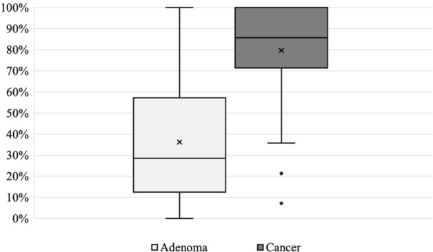
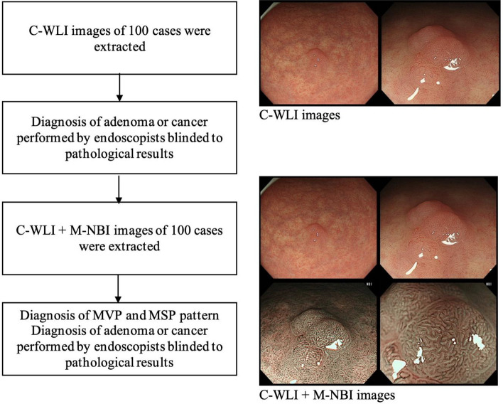
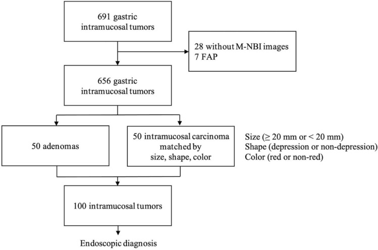
This study assessed the effect of magnifying endoscopy with narrow-band imaging (M-NBI) on the endoscopic differential diagnosis between intramucosal gastric carcinomas and adenomas with matched characteristics. Associations between magnified endoscopic findings and pathological high-grade cellular and architectural atypia were also investigated. In total, the records of 50 adenomas and 50 intramucosal well-differentiated adenocarcinomas matched by tumor size (≥ 20 mm or < 20 mm), shape (depression or non-depression), and color (red or non-red) were extracted. Fourteen endoscopists diagnosed adenoma or cancer in the 100 cases with conventional white light imaging (C-WLI), then did the same with C-WLI + M-NBI.The cancer diagnostic sensitivity, specificity, and accuracy were assessed. The sensitivity of C-WLI + M-NBI for cancer diagnosis was 79.9% compared to 71.6% with C-WLI (p < 0.001). There were no significant differences in specificity (40.1% vs. 36.3%, p = 0.296) and accuracy (55.9% vs. 58.1%, p = 0.163). High-grade cytological or architectural atypia was diagnosed more often with irregular microvascular pattern (IMVP) or microsurface pattern (IMSP), respectively, than the low-grade forms. In conclusion, IMVP and IMSP correlate with high-grade cytological and architectural atypia. M-NBI is useful in differentiating intramucosal carcinoma from adenoma and can reduce underdiagnosis of cancer.
本研究评估了窄带成像放大内镜(M-NBI)对大小(≥20mm 或<20mm)、形态(凹陷或非凹陷)和颜色(红色或非红色)匹配的黏膜内胃癌和腺瘤的内镜鉴别诊断的影响。还研究了放大内镜下表现与高等级细胞和结构异型性的病理之间的关系。共提取了 50 个腺瘤和 50 个大小(≥20mm 或<20mm)、形态(凹陷或非凹陷)和颜色(红色或非红色)匹配的黏膜内高分化腺癌的记录。14 位内镜医师在 100 例常规白光成像(C-WLI)中诊断腺瘤或癌症,然后用 C-WLI+M-NBI 进行相同的诊断。评估了癌症诊断的敏感性、特异性和准确性。C-WLI+M-NBI 对癌症诊断的敏感性为 79.9%,而 C-WLI 为 71.6%(p<0.001)。特异性(40.1%比 36.3%,p=0.296)和准确性(55.9%比 58.1%,p=0.163)无显著差异。不规则微血管模式(IMVP)或微表面模式(IMSP)的高等级细胞学或结构异型性比低等级形式更常被诊断。总之,IMVP 和 IMSP 与高等级细胞学和结构异型性相关。M-NBI 有助于区分黏膜内癌和腺瘤,并可减少癌症的漏诊。