Department of Radiology, Peking Union Medical College Hospital, Peking Union Medical College & Chinese Academy of Medical Sciences, Beijing, 100730, China.
CAS Key Laboratory of Molecular Imaging, Beijing Key Laboratory of Molecular Imaging, the State Key Laboratory of Management and Control for Complex Systems, Institute of Automation, Chinese Academy of Sciences, Beijing, 100190, China.
Eur J Nucl Med Mol Imaging. 2022 Jul;49(8):2723-2734. doi: 10.1007/s00259-022-05834-5. Epub 2022 May 20.
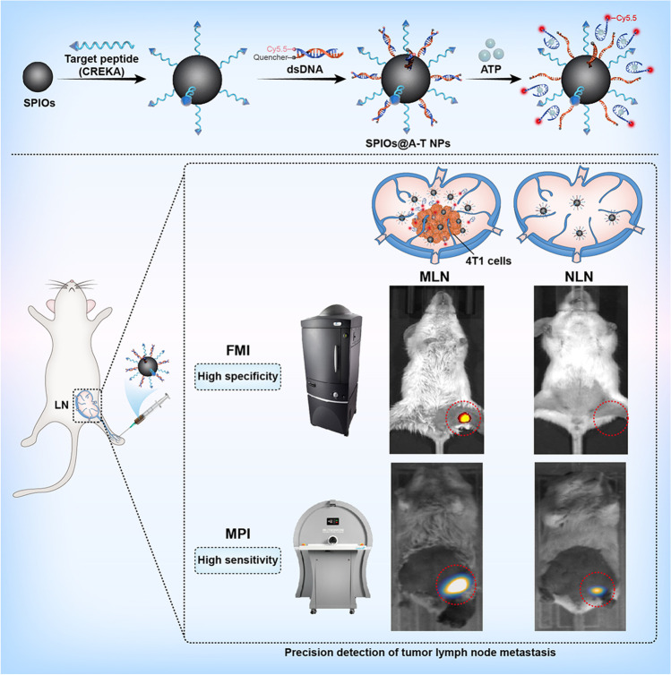
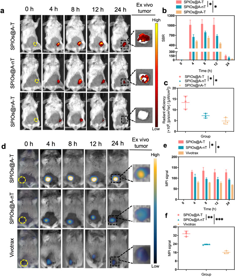
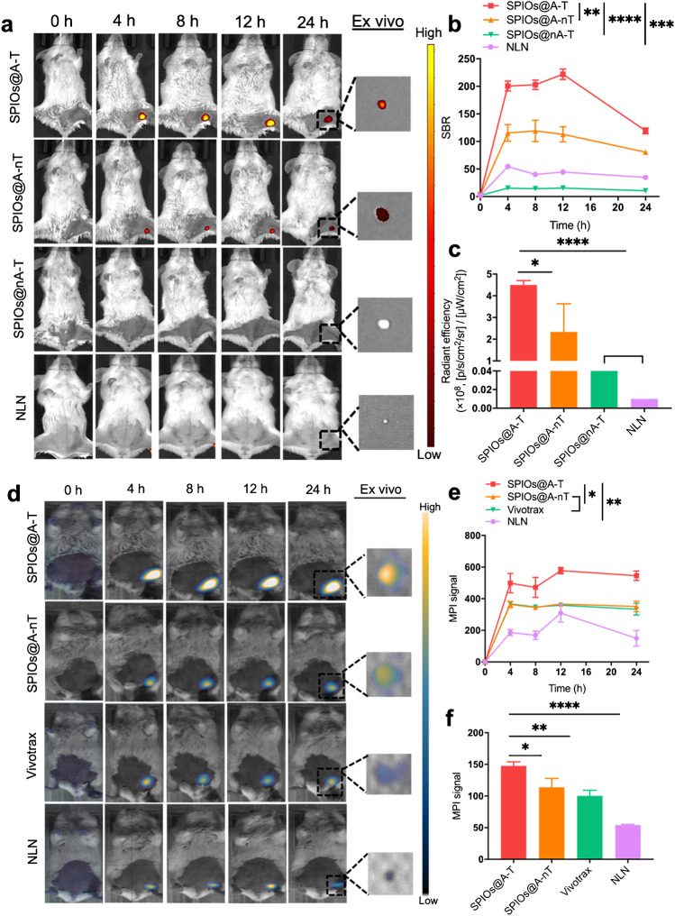
A sensitive and specific imaging method to detect metastatic cancer cells in lymph nodes to detect the early-stage breast cancer is still a challenge. The purpose of this study was to investigate a novel breast cancer-targeting and tumour microenvironment ATP-responsive superparamagnetic iron oxide nanoparticles (SPIOs) imaging probe (abbreviated as SPIOs@A-T) that was developed to detect lymph node metastasis through fluorescence molecular imaging (FMI) and magnetic particle imaging (MPI).
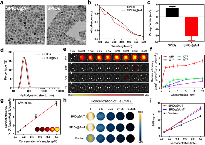
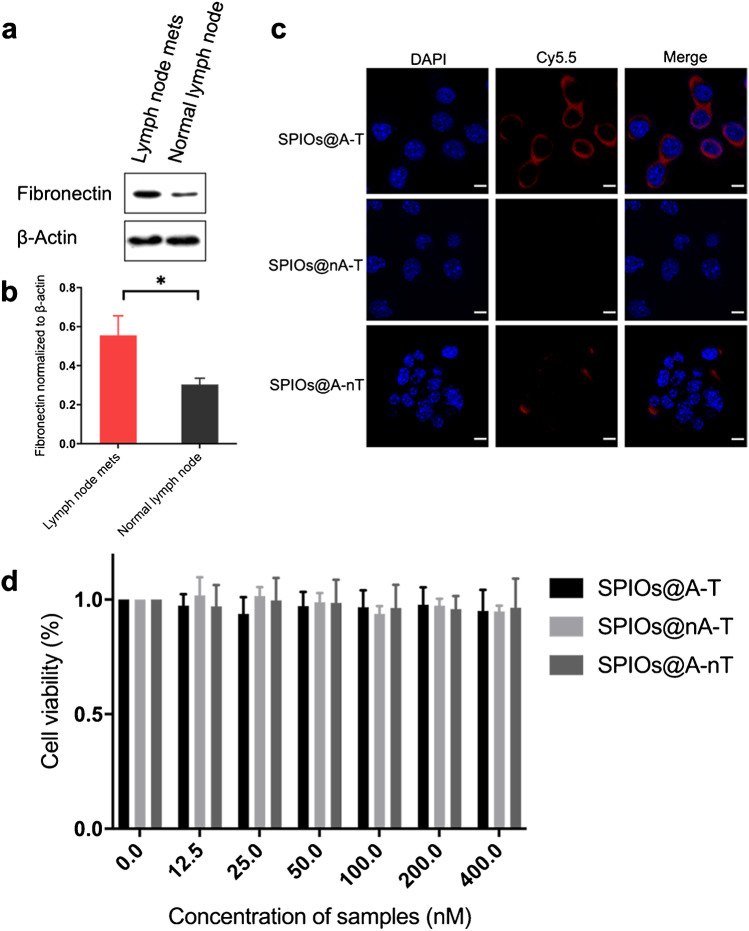
The conjugation of the targeted peptide CREKA and SPIOs was via linker sulfo-SMCC, while the dsDNA-Cy5.5 was modified on SPIOs through the conjugation between maleimide group in sulfo-SMCC and sulfydryl group in dsDNA-Cy5.5. SPIOs@A-T was characterised for its imaging properties, targeting ability and toxicity in vitro. Mice with metastatic lymph node (MLN) of breast cancer were established to evaluate the FMI and MPI imaging strategy in vivo. Healthy mice with normal lymph node (NLN) were used as control group. Histological examination and biosafety evaluation were performed for further assessment.
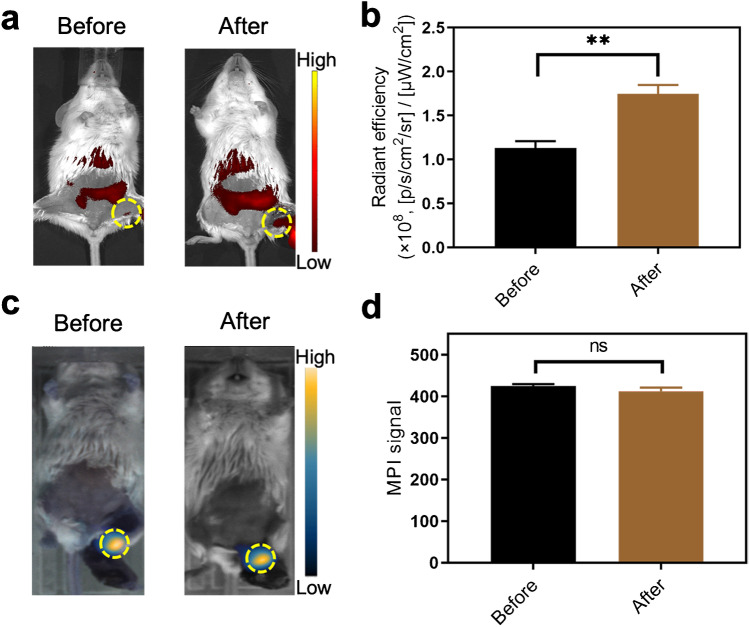
After injection with SPIOs@A-T, the obvious high fluorescent intensity and MPI signal were observed in MLN group than those in NLN group. FMI can specifically light up MLN using an ATP-responsive fluorescence design. On the other hand, MPI could complement the limitation of imaging depth from FMI and could detect MLN more sensitively. Besides, the biosafety evaluation results showed SPIOs@A-T had no detectable biological toxicity.
SPIOs@A-T imaging probe in combination with FMI and MPI can provide a promising novel method for the precise detection of MLN in vivo.
寻找一种灵敏且特异的方法来检测淋巴结中的转移性癌细胞,以发现早期乳腺癌,这仍然是一个挑战。本研究旨在探索一种新型的乳腺癌靶向和肿瘤微环境 ATP 响应超顺磁性氧化铁纳米粒子(SPIOs)成像探针(简称 SPIOs@A-T),该探针通过荧光分子成像(FMI)和磁性粒子成像(MPI)来检测淋巴结转移。
通过连接子 sulfo-SMCC 将靶向肽 CREKA 与 SPIOs 偶联,而 dsDNA-Cy5.5 通过 sulfo-SMCC 中的马来酰亚胺基团和 dsDNA-Cy5.5 中的巯基之间的共轭修饰在 SPIOs 上。对 SPIOs@A-T 的成像特性、靶向能力和体外毒性进行了表征。建立了具有转移性淋巴结(MLN)的乳腺癌小鼠模型,以评估体内 FMI 和 MPI 成像策略。将具有正常淋巴结(NLN)的健康小鼠用作对照组。进行了组织学检查和生物安全性评估,以进一步评估。
注射 SPIOs@A-T 后,MLN 组的荧光强度和 MPI 信号明显高于 NLN 组。FMI 可以使用 ATP 响应荧光设计特异性地点亮 MLN。另一方面,MPI 可以弥补 FMI 成像深度的限制,并且可以更灵敏地检测 MLN。此外,生物安全性评估结果表明 SPIOs@A-T 没有可检测的生物毒性。
SPIOs@A-T 成像探针结合 FMI 和 MPI 可为体内 MLN 的精确检测提供一种有前途的新方法。