• 文献检索
  • 文档翻译
  • 深度研究
  • 学术资讯
  • Suppr Zotero 插件Zotero 插件
  • 邀请有礼
  • 套餐&价格
  • 历史记录
应用&插件
Suppr Zotero 插件Zotero 插件浏览器插件Mac 客户端Windows 客户端微信小程序
定价
高级版会员购买积分包购买API积分包
服务
文献检索文档翻译深度研究API 文档MCP 服务
关于我们
关于 Suppr公司介绍联系我们用户协议隐私条款
关注我们

Suppr 超能文献

核心技术专利:CN118964589B侵权必究
粤ICP备2023148730 号-1Suppr @ 2026

文献检索

告别复杂PubMed语法,用中文像聊天一样搜索,搜遍4000万医学文献。AI智能推荐,让科研检索更轻松。

立即免费搜索

文件翻译

保留排版,准确专业,支持PDF/Word/PPT等文件格式,支持 12+语言互译。

免费翻译文档

深度研究

AI帮你快速写综述,25分钟生成高质量综述,智能提取关键信息,辅助科研写作。

立即免费体验

基于 CT1258 前列腺癌体内模型,用氯化锰(MnCl2)和超顺磁氧化铁纳米颗粒(SPIOs)进行纵向 MRI 对比增强监测早期肿瘤发展。

Longitudinal MRI contrast enhanced monitoring of early tumour development with manganese chloride (MnCl2) and superparamagnetic iron oxide nanoparticles (SPIOs) in a CT1258 based in vivo model of prostate cancer.

机构信息

University of Veterinary Medicine Hannover, Hannover, Germany.

出版信息

BMC Cancer. 2012 Jul 11;12:284. doi: 10.1186/1471-2407-12-284.

DOI:10.1186/1471-2407-12-284
PMID:22784304
原文链接:https://pmc.ncbi.nlm.nih.gov/articles/PMC3520113/
Abstract

BACKGROUND

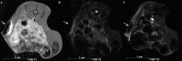
Cell lines represent a key tool in cancer research allowing the generation of neoplasias which resemble initial tumours in in-vivo animal models. The characterisation of early tumour development is of major interest in order to evaluate the efficacy of therapeutic agents. Magnetic resonance imaging (MRI) based in-vivo characterisation allows visualisation and characterisation of tumour development in early stages prior to manual palpation. Contrast agents for MRI such as superparamagnetic iron oxide nanoparticles (SPIOs) and manganese chloride (MnCl2) represent powerful tools for the in-vivo characterisation of early stage tumours. In this experimental study, we labelled prostate cancer cells with MnCl2 or SPIOs in vitro and used 1 T MRI for tracing labelled cells in-vitro and 7 T MRI for tracking in an in-vivo animal model.

METHODS

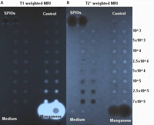
Labelling of prostate cancer cells CT1258 was established in-vitro with MnCl2 and SPIOs. In-vitro detection of labelled cells in an agar phantom was carried out through 1 T MRI while in-vivo detection was performed using 7 T MRI after subcutaneous (s.c.) injection of labelled cells into NOD-Scid mice (n = 20). The animals were scanned in regular intervals until euthanization. The respective tumour volumes were analysed and corresponding tumour masses were subjected to histologic examination.

RESULTS

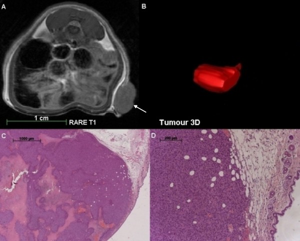
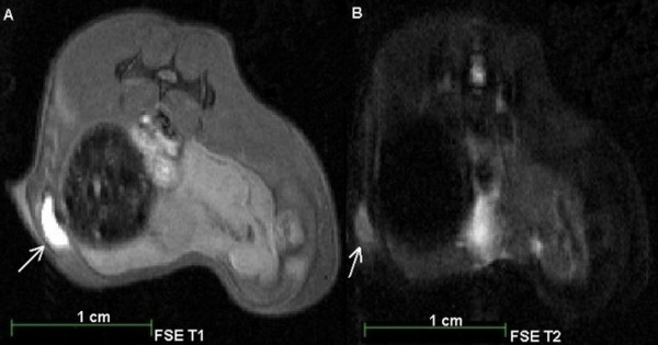
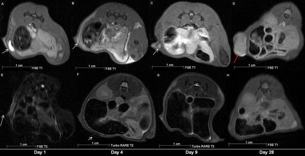
MnCl2in-vitro labelling resulted in no significant metabolic effects on proliferation and cell vitality. In-vitro detection-limit accounted 105 cells for MnCl2 as well as for SPIOs labelling. In-vivo 7 T MRI scans allowed detection of 103 and 104 cells. In-vivo MnCl2 labelled cells were detectable from days 4-16 while SPIO labelling allowed detection until 4 days after s.c. injection. MnCl2 labelled cells were highly tumourigenic in NOD-Scid mice and the tumour volume development was characterised in a time dependent manner. The amount of injected cells correlated with tumour size development and disease progression. Histological analysis of the induced tumour masses demonstrated characteristic morphologies of prostate adenocarcinoma.

CONCLUSIONS

To the best of our knowledge, this is the first study reporting direct in-vitro MnCl2 labelling and 7 T based in-vivo MRI tracing of cancer cells in a model of prostate cancer. MnCl2 labelling was found to be suitable for in-vivo tracing allowing long detection periods. The labelled cells kept their highly tumourigenic potential in-vivo. Tumour volume development was visualised prior to manual palpation allowing tumour characterisation in early stages of the disease.

摘要

背景

细胞系是癌症研究的重要工具,可生成类似于体内动物模型中初始肿瘤的肿瘤。早期肿瘤发展的特征是评估治疗药物疗效的主要关注点。基于磁共振成像(MRI)的体内特征可以在手动触诊之前可视化和表征早期肿瘤的发展。MRI 用的造影剂,如超顺磁氧化铁纳米颗粒(SPIOs)和氯化锰(MnCl2),是用于早期肿瘤体内特征的有力工具。在这项实验研究中,我们在体外用 MnCl2 或 SPIOs 标记前列腺癌细胞,并用 1T MRI 进行体外标记细胞的跟踪,用 7T MRI 进行体内动物模型的跟踪。

方法

在体外用 MnCl2 和 SPIOs 对前列腺癌细胞 CT1258 进行标记。通过 1T MRI 在琼脂体模中对标记细胞进行体外检测,通过皮下(s.c.)注射标记细胞到 NOD-Scid 小鼠后,用 7T MRI 进行体内检测(n=20)。动物每隔一段时间扫描,直到安乐死。分析相应的肿瘤体积,并对相应的肿瘤质量进行组织学检查。

结果

MnCl2 体外标记对增殖和细胞活力没有显著的代谢影响。体外检测限为 105 个细胞,MnCl2 和 SPIOs 标记的细胞都可以检测到。体内 7T MRI 扫描可以检测到 103 和 104 个细胞。体内 MnCl2 标记的细胞可在第 4-16 天检测到,而 SPIO 标记的细胞可在注射后第 4 天检测到。MnCl2 标记的细胞在 NOD-Scid 小鼠中具有很强的致瘤性,肿瘤体积的发展呈时间依赖性。注射细胞的数量与肿瘤大小的发展和疾病的进展相关。诱导的肿瘤组织学分析显示出前列腺腺癌的特征形态。

结论

据我们所知,这是首例报道直接在体外使用 MnCl2 标记和在体内使用 7T 基于 MRI 跟踪前列腺癌模型中癌细胞的研究。MnCl2 标记被发现适合于体内跟踪,允许长时间的检测。标记的细胞在体内保持其高度致瘤性。在手动触诊之前,可以观察到肿瘤体积的发展,从而在疾病的早期阶段对肿瘤进行特征化。

https://cdn.ncbi.nlm.nih.gov/pmc/blobs/ee50/3520113/6758290881f6/1471-2407-12-284-5.jpg
https://cdn.ncbi.nlm.nih.gov/pmc/blobs/ee50/3520113/4edeeefacae9/1471-2407-12-284-1.jpg
https://cdn.ncbi.nlm.nih.gov/pmc/blobs/ee50/3520113/6ea5d83aeb04/1471-2407-12-284-2.jpg
https://cdn.ncbi.nlm.nih.gov/pmc/blobs/ee50/3520113/f7894cdf517d/1471-2407-12-284-3.jpg
https://cdn.ncbi.nlm.nih.gov/pmc/blobs/ee50/3520113/ab55d0499681/1471-2407-12-284-4.jpg
https://cdn.ncbi.nlm.nih.gov/pmc/blobs/ee50/3520113/6758290881f6/1471-2407-12-284-5.jpg

相似文献

1
Longitudinal MRI contrast enhanced monitoring of early tumour development with manganese chloride (MnCl2) and superparamagnetic iron oxide nanoparticles (SPIOs) in a CT1258 based in vivo model of prostate cancer.基于 CT1258 前列腺癌体内模型,用氯化锰(MnCl2)和超顺磁氧化铁纳米颗粒(SPIOs)进行纵向 MRI 对比增强监测早期肿瘤发展。
BMC Cancer. 2012 Jul 11;12:284. doi: 10.1186/1471-2407-12-284.
2
Monitoring of in vivo function of superparamagnetic iron oxide labelled murine dendritic cells during anti-tumour vaccination.在抗肿瘤疫苗接种过程中监测超顺磁性氧化铁标记的鼠树突状细胞的体内功能。
PLoS One. 2011;6(5):e19662. doi: 10.1371/journal.pone.0019662. Epub 2011 May 27.
3
Imaging monocytes with iron oxide nanoparticles targeted towards the monocyte integrin MAC-1 (CD11b/CD18) does not result in improved atherosclerotic plaque detection by in vivo MRI.用氧化铁纳米颗粒对单核细胞整合素 MAC-1(CD11b/CD18)进行靶向成像,并不会通过体内 MRI 提高动脉粥样硬化斑块的检测效果。
Contrast Media Mol Imaging. 2010 Sep-Oct;5(5):268-75. doi: 10.1002/cmmi.384.
4
Current limitations of molecular magnetic resonance imaging for tumors as evaluated with high-relaxivity CD105-specific iron oxide nanoparticles.用高弛豫率 CD105 特异性氧化铁纳米颗粒评估肿瘤的分子磁共振成像的当前局限性。
Invest Radiol. 2012 Jul;47(7):383-91. doi: 10.1097/RLI.0b013e31824c5a57.
5
PSMA-targeting iron oxide magnetic nanoparticles enhance MRI of preclinical prostate cancer.靶向前列腺特异性膜抗原的氧化铁磁性纳米颗粒增强临床前前列腺癌的磁共振成像。
Nanomedicine (Lond). 2015 Feb;10(3):375-86. doi: 10.2217/nnm.14.122. Epub 2014 Nov 19.
6
Quantitative Activity Measurements of Brown Adipose Tissue at 7 T Magnetic Resonance Imaging After Application of Triglyceride-Rich Lipoprotein 59Fe-Superparamagnetic Iron Oxide Nanoparticle: Intravenous Versus Intraperitoneal Approach.富含甘油三酯的脂蛋白59Fe-超顺磁性氧化铁纳米颗粒应用后7T磁共振成像对棕色脂肪组织的定量活性测量:静脉注射与腹腔注射途径
Invest Radiol. 2016 Mar;51(3):194-202. doi: 10.1097/RLI.0000000000000235.
7
In vivo MRI of intraspinally injected SPIO-labelled human CD34+ cells in a transgenic mouse model of ALS.体内 MRI 示踪转染 SPIO 标记的人 CD34+细胞在肌萎缩侧索硬化症转基因小鼠模型中的应用。
In Vivo. 2012 Jan-Feb;26(1):31-8.
8
Establishing an in vivo model of canine prostate carcinoma using the new cell line CT1258.利用新细胞系CT1258建立犬前列腺癌体内模型。
BMC Cancer. 2008 Aug 15;8:240. doi: 10.1186/1471-2407-8-240.
9
Formulation of novel lipid-coated magnetic nanoparticles as the probe for in vivo imaging.新型脂质包覆磁性纳米粒子的制剂作为体内成像的探针。
J Biomed Sci. 2009 Sep 21;16(1):86. doi: 10.1186/1423-0127-16-86.
10
In vivo visualization and ex vivo quantification of murine breast cancer cells in the mouse brain using MRI cell tracking and electron paramagnetic resonance.利用磁共振成像细胞追踪和电子顺磁共振对小鼠大脑中的小鼠乳腺癌细胞进行体内可视化和体外定量分析。
NMR Biomed. 2015 Mar;28(3):367-75. doi: 10.1002/nbm.3259. Epub 2015 Jan 22.

引用本文的文献

1
The role of manganese-based MRI contrast agents for cancer theranostics: Where do we stand in 2025?锰基磁共振成像造影剂在癌症诊疗中的作用:到2025年我们处于什么水平?
Theranostics. 2025 Mar 15;15(9):4147-4174. doi: 10.7150/thno.108705. eCollection 2025.
2
Manganese-Based Nanotheranostics for Magnetic Resonance Imaging-Mediated Precise Cancer Management.基于锰的纳米诊疗剂用于磁共振成像介导的精确癌症管理。
Int J Nanomedicine. 2023 Oct 26;18:6077-6099. doi: 10.2147/IJN.S426311. eCollection 2023.
3
A primer on cell tracking using MRI.磁共振成像细胞追踪入门

本文引用的文献

1
Development of a brain metastatic canine prostate cancer cell line.建立脑转移犬前列腺癌细胞系。
Prostate. 2011 Sep;71(12):1251-63. doi: 10.1002/pros.21341. Epub 2011 Feb 14.
2
Global cancer statistics.全球癌症统计数据。
CA Cancer J Clin. 2011 Mar-Apr;61(2):69-90. doi: 10.3322/caac.20107. Epub 2011 Feb 4.
3
Formulation of novel lipid-coated magnetic nanoparticles as the probe for in vivo imaging.新型脂质包覆磁性纳米粒子的制剂作为体内成像的探针。
Front Med (Lausanne). 2023 May 31;10:1193459. doi: 10.3389/fmed.2023.1193459. eCollection 2023.
4
Establishment and characterization of stable red, far-red (fR) and near infra-red (NIR) transfected canine prostate cancer cell lines.稳定转染红色、远红光(fR)和近红外光(NIR)的犬前列腺癌细胞系的建立与表征
Cancer Cell Int. 2020 Apr 29;20:139. doi: 10.1186/s12935-020-01211-0. eCollection 2020.
5
A brief review of cytotoxicity of nanoparticles on mesenchymal stem cells in regenerative medicine.纳米颗粒对再生医学中间充质干细胞细胞毒性的简要综述。
Int J Nanomedicine. 2019 May 24;14:3875-3892. doi: 10.2147/IJN.S205574. eCollection 2019.
6
Genetic engineered molecular imaging probes for applications in cell therapy: emphasis on MRI approach.用于细胞治疗的基因工程分子成像探针:重点介绍磁共振成像方法
Am J Nucl Med Mol Imaging. 2016 Sep 22;6(5):234-261. eCollection 2016.
7
Nanoparticles and clinically applicable cell tracking.纳米颗粒与临床适用的细胞追踪
Br J Radiol. 2015 Oct;88(1054):20150375. doi: 10.1259/bjr.20150375. Epub 2015 Aug 7.
8
MRI tracking stem cells transplantation for coronary heart disease.MRI 示踪干细胞移植治疗冠心病。
Pak J Med Sci. 2014 Jul;30(4):899-903. doi: 10.12669/pjms.304.4936.
9
Generation and characterisation of a canine EGFP-HMGA2 prostate cancer in vitro model.犬源EGFP-HMGA2前列腺癌体外模型的构建与表征
PLoS One. 2014 Jun 10;9(6):e98788. doi: 10.1371/journal.pone.0098788. eCollection 2014.
J Biomed Sci. 2009 Sep 21;16(1):86. doi: 10.1186/1423-0127-16-86.
4
Labelling of mammalian cells for visualisation by MRI.用于 MRI 可视化的哺乳动物细胞标记。
Eur Radiol. 2010 Feb;20(2):255-74. doi: 10.1007/s00330-009-1540-1. Epub 2009 Aug 12.
5
Magnetic resonance imaging of human embryonic stem cells.人类胚胎干细胞的磁共振成像
Curr Protoc Stem Cell Biol. 2009 Aug;Chapter 5:Unit 5A.3. doi: 10.1002/9780470151808.sc05a03s10.
6
In vivo MRI cell tracking: clinical studies.体内磁共振成像细胞追踪:临床研究
AJR Am J Roentgenol. 2009 Aug;193(2):314-25. doi: 10.2214/AJR.09.3107.
7
Manganese-guided cellular MRI of human embryonic stem cell and human bone marrow stromal cell viability.锰引导的人胚胎干细胞和人骨髓基质细胞活力的细胞 MRI。
Magn Reson Med. 2009 Oct;62(4):1047-54. doi: 10.1002/mrm.22071.
8
Cell labeling and tracking for experimental models using magnetic resonance imaging.使用磁共振成像对实验模型进行细胞标记和追踪。
Methods. 2009 Jun;48(2):112-24. doi: 10.1016/j.ymeth.2009.03.020. Epub 2009 Apr 9.
9
In vitro labeling of human embryonic stem cells for magnetic resonance imaging.用于磁共振成像的人胚胎干细胞的体外标记
J Vis Exp. 2008 Aug 3(17):827. doi: 10.3791/827.
10
Detection and quantification of magnetically labeled cells by cellular MRI.通过细胞磁共振成像检测和定量磁性标记的细胞。
Eur J Radiol. 2009 May;70(2):258-64. doi: 10.1016/j.ejrad.2008.09.021. Epub 2008 Nov 7.