Department of Leukemia, The University of Texas MD Anderson Cancer Center, Houston, TX.
Division of Hematology, Davidoff Cancer Center, Rabin Medical Center, Petah Tikva, Israel; and.
J Immunol. 2022 Jun 15;208(12):2847-2855. doi: 10.4049/jimmunol.2101105. Epub 2022 May 20.
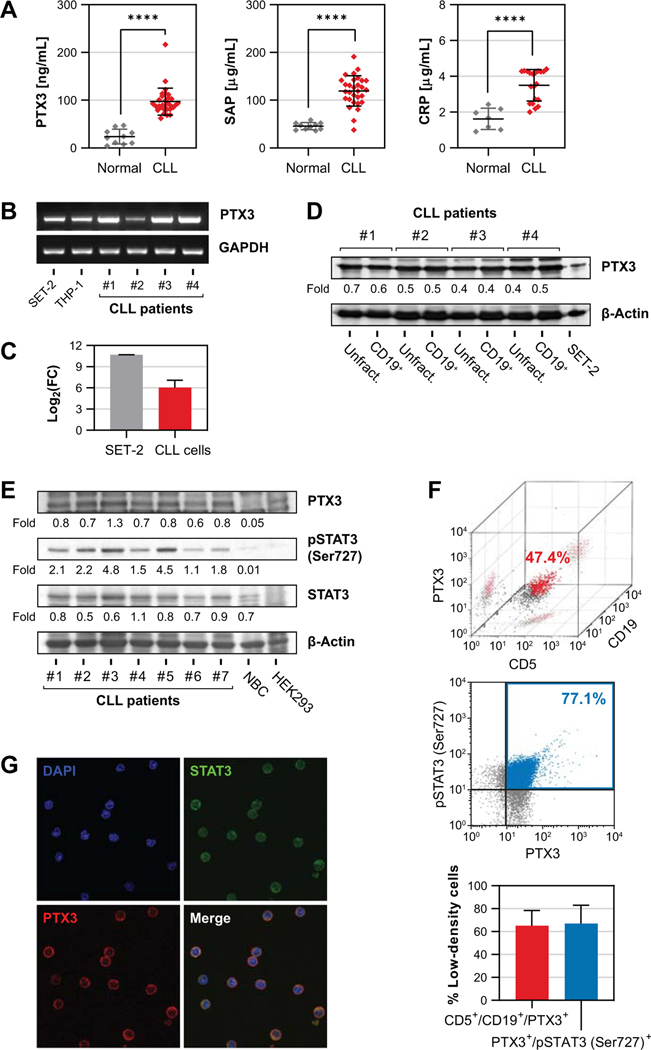
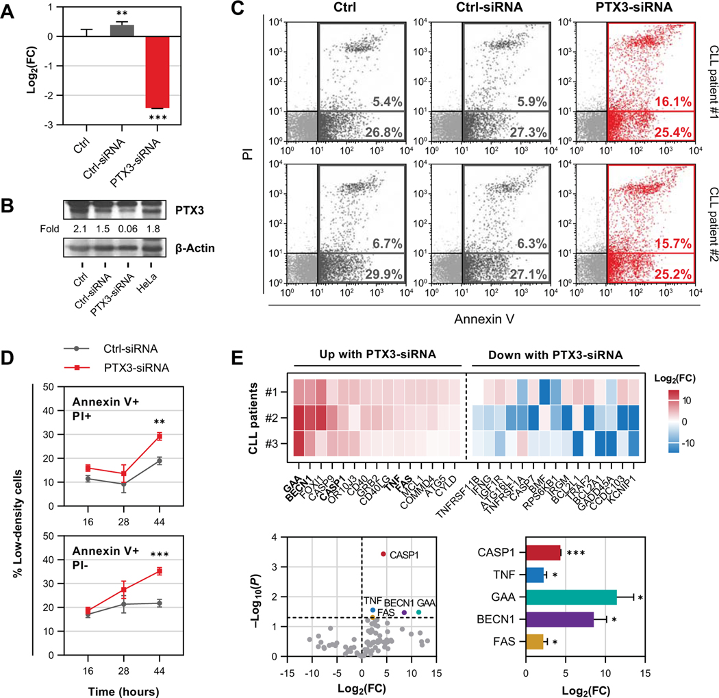
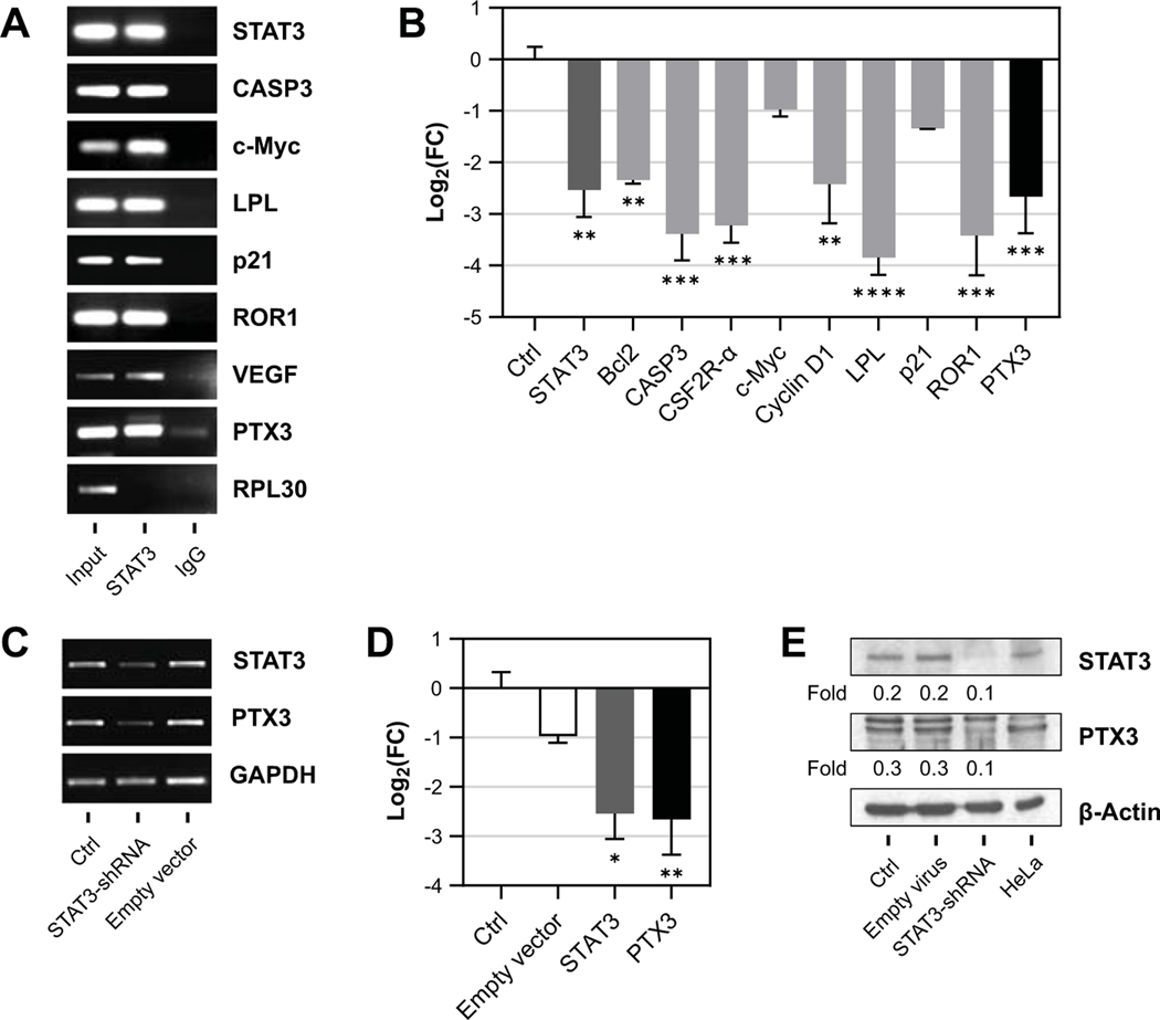
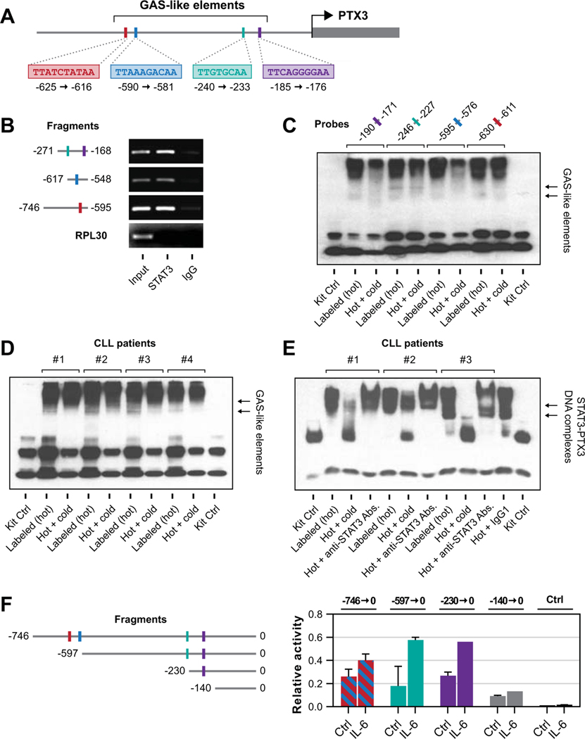
Pentraxin-related protein 3 (PTX3), commonly produced by myeloid and endothelial cells, is a humoral pattern recognition protein of the innate immune system. Because PTX3 plasma levels of patients with chronic lymphocytic leukemia (CLL) are high and most circulating cells in patients with CLL are CLL cells, we reasoned that CLL cells produce PTX3. Western immunoblotting revealed that low-density cells from seven of seven patients with CLL produce high levels of PTX3, flow cytometry analysis revealed that the PTX3-producing cells are B lymphocytes coexpressing CD19 and CD5, and confocal microscopy showed that PTX3 is present in the cytoplasm of CLL cells. Because STAT3 is constitutively activated in CLL cells, and because we identified putative STAT3 binding sites within the PTX3 gene promoter, we postulated that phosphorylated STAT3 triggers transcriptional activation of PTX3. Immunoprecipitation analysis of CLL cells' chromatin fragments showed that STAT3 Abs precipitated PTX3 DNA. STAT3 knockdown induced a marked reduction in PTX3 expression, indicating a STAT3-induced transcriptional activation of the PTX3 gene in CLL cells. Using an EMSA, we established and used a dual-reporter luciferase assay to confirm that STAT3 binds the PTX3 gene promoter. Downregulation of PTX3 enhanced apoptosis of CLL cells, suggesting that inhibition of PTX3 might benefit patients with CLL.
血清淀粉样蛋白相关蛋白 3(PTX3),通常由髓样细胞和内皮细胞产生,是先天免疫系统的一种体液模式识别蛋白。由于慢性淋巴细胞白血病(CLL)患者的 PTX3 血浆水平较高,并且 CLL 患者的大多数循环细胞都是 CLL 细胞,因此我们推断 CLL 细胞产生 PTX3。Western 免疫印迹显示,7 例 CLL 患者的低密度细胞产生高水平的 PTX3,流式细胞术分析显示,产生 PTX3 的细胞是共表达 CD19 和 CD5 的 B 淋巴细胞,共聚焦显微镜显示 PTX3 存在于 CLL 细胞的细胞质中。由于 STAT3 在 CLL 细胞中持续激活,并且我们在 PTX3 基因启动子内鉴定出了假定的 STAT3 结合位点,因此我们假设磷酸化的 STAT3 触发 PTX3 的转录激活。CLL 细胞染色质片段的免疫沉淀分析表明,STAT3 Abs 沉淀了 PTX3 DNA。STAT3 敲低诱导了 PTX3 表达的显著减少,表明 STAT3 在 CLL 细胞中诱导了 PTX3 基因的转录激活。通过 EMSA,我们建立并使用双报告荧光素酶测定法证实 STAT3 结合 PTX3 基因启动子。下调 PTX3 增强了 CLL 细胞的凋亡,这表明抑制 PTX3 可能有益于 CLL 患者。